
 RPS3 polyclonal antibody
  Datasheet
 COA
COA MSDS
MSDS SHIP
SHIP
Product Name :
RPS3 polyclonal antibody Background :
Ribosomes, the organelles that catalyze protein synthesis, consist of a small 40S subunit and a large 60S subunit. Together these subunits are composed of 4 RNA species and approximately 80 structurally distinct proteins. This gene encodes a ribosomal protein that is a component of the 40S subunit, where it forms part of the domain where translation is initiated. The protein belongs to the S3P family of ribosomal proteins. Studies of the mouse and rat proteins have demonstrated that the protein has an extraribosomal role as an endonuclease involved in the repair of UV-induced DNA damage. The protein appears to be located in both the cytoplasm and nucleus but not in the nucleolus. Higher levels of expression of this gene in colon adenocarcinomas and adenomatous polyps compared to adjacent normal colonic mucosa have been observed. This gene is co-transcribed with the small nucleolar RNA genes U15A and U15B, which are located in its first and fifth introns, respectively. As is typical for genes encoding ribosomal proteins, there are multiple processed pseudogenes of this gene dispersed through the genome. Multiple alternatively spliced transcript variants encoding different isoforms have been found for this gene. Product :
1mg/ml in PBS with 0.02% sodium azide, 50% glycerol, pH7.2 Storage&Stability :
Store at 4°C short term. Aliquot and store at -20°C long term. Avoid freeze-thaw cycles. Specificity :
Unmodification Immunogen :
Recombinant fusion protein of human RPS3(NP_000996.2). Conjugate :
Unconjugated Modification :
Unmodification
-
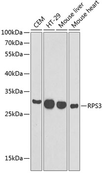 Western blot analysis of extracts of various cell lines, using RPS3 antibody at 1:1000 dilution.
Secondary antibody: HRP Goat Anti-Rabbit IgG at 1:10000 dilution.
Lysates/proteins: 25ug per lane.
Blocking buffer: 3% nonfat dry milk in TBST. -
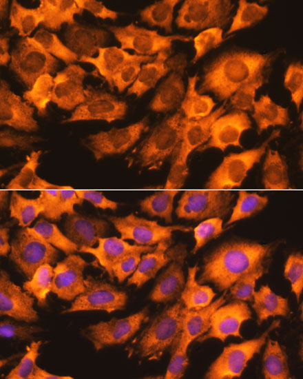 Immunofluorescence analysis of C6 cells using RPS3 antibody at dilution of 1:100. Blue: DAPI for nuclear staining.
 -
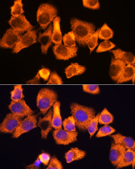 Immunofluorescence analysis of C6 cells using RPS3 antibody at dilution of 1:100. Blue: DAPI for nuclear staining.
 -
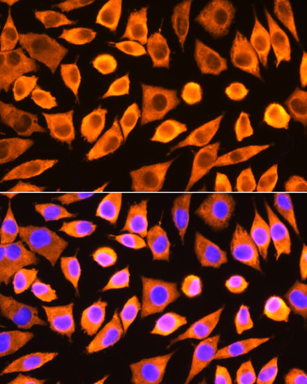 Immunofluorescence analysis of C6 cells using RPS3 antibody at dilution of 1:100. Blue: DAPI for nuclear staining.
Bioworld Biotech only provide peptides for our antibodies and do not provide additional peptide customization services.
Price/Size :
USD 368/1mg/vial

Tips:
For phospho antibody, we provide phospho peptide(0.5mg) and non-phospho peptide(0.5mg).Describe :
Blocking peptides are peptides that bind specifically to the target antibody and block antibody binding. These peptide usually contains the epitope recognized by the antibody. Antibodies bound to the blocking peptide no longer bind to the epitope on the target protein. This mechanism is useful when non-specific binding is an issue, for example, in Western blotting (WB) and Immunohistochemistry (IHC). By comparing the staining from the blocked antibody versus the antibody alone, one can see which staining is specific; Specific binding will be absent from the western blot or IHC performed with the neutralized antibody.Formula:
Synthetic peptide was lyophilized with 100% acetonitrile and is supplied as a powder. Reconstitute with 0.1 ml DI water for a final concentration of 10 mg/ml.The purity is >90%,tested by HPLC and MS.
Storage:
The freeze-dried powder is more stable. For short time at 2-8°C. For long term storage store at -20°C.
Note :
This product is for research use only (RUO only). Not for use in diagnostic or therapeutic procedures.