
 TNFRSF25 polyclonal antibody
  Datasheet
 COA
COA MSDS
MSDS SHIP
SHIP
Product Name :
TNFRSF25 polyclonal antibody Background :
The protein encoded by this gene is a member of the TNF-receptor superfamily. This receptor is expressed preferentially in the tissues enriched in lymphocytes, and it may play a role in regulating lymphocyte homeostasis. This receptor has been shown to stimulate NF-kappa B activity and regulate cell apoptosis. The signal transduction of this receptor is mediated by various death domain containing adaptor proteins. Knockout studies in mice suggested the role of this gene in the removal of self-reactive T cells in the thymus. Multiple alternatively spliced transcript variants of this gene encoding distinct isoforms have been reported, most of which are potentially secreted molecules. The alternative splicing of this gene in B and T cells encounters a programmed change upon T-cell activation, which predominantly produces full-length, membrane bound isoforms, and is thought to be involved in controlling lymphocyte proliferation induced by T-cell activation. Product :
1mg/ml in PBS with 0.02% sodium azide, 50% glycerol, pH7.2 Storage&Stability :
Store at 4°C short term. Aliquot and store at -20°C long term. Avoid freeze-thaw cycles. Specificity :
Unmodification Immunogen :
Recombinant fusion protein of human TNFRSF25(NP_683866.1). Conjugate :
Unconjugated Modification :
Unmodification
-
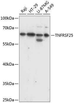 Western blot analysis of extracts of various cell lines, using TNFRSF25 antibody at 1:1000 dilution.
Secondary antibody: HRP Goat Anti-Rabbit IgG at 1:10000 dilution.
Lysates/proteins: 25ug per lane.
Blocking buffer: 3% nonfat dry milk in TBST.
Detection: ECL Basic Kit .
Exposure time: 60s. -
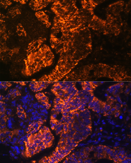 Immunofluorescence analysis of human colon carcinoma cells using TNFRSF25 antibody at dilution of 1:100. Blue: DAPI for nuclear staining.
 -
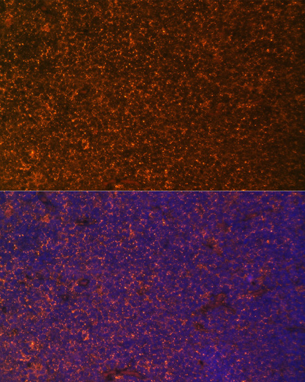 Immunofluorescence analysis of human colon carcinoma cells using TNFRSF25 antibody at dilution of 1:100. Blue: DAPI for nuclear staining.
Bioworld Biotech only provide peptides for our antibodies and do not provide additional peptide customization services.
Price/Size :
USD 368/1mg/vial

Tips:
For phospho antibody, we provide phospho peptide(0.5mg) and non-phospho peptide(0.5mg).Describe :
Blocking peptides are peptides that bind specifically to the target antibody and block antibody binding. These peptide usually contains the epitope recognized by the antibody. Antibodies bound to the blocking peptide no longer bind to the epitope on the target protein. This mechanism is useful when non-specific binding is an issue, for example, in Western blotting (WB) and Immunohistochemistry (IHC). By comparing the staining from the blocked antibody versus the antibody alone, one can see which staining is specific; Specific binding will be absent from the western blot or IHC performed with the neutralized antibody.Formula:
Synthetic peptide was lyophilized with 100% acetonitrile and is supplied as a powder. Reconstitute with 0.1 ml DI water for a final concentration of 10 mg/ml.The purity is >90%,tested by HPLC and MS.
Storage:
The freeze-dried powder is more stable. For short time at 2-8°C. For long term storage store at -20°C.
Note :
This product is for research use only (RUO only). Not for use in diagnostic or therapeutic procedures.