
 IRF3 (Phospho-S396) polyclonal antibody
  Datasheet
 COA
COA MSDS
MSDS SHIP
SHIP
Product Name :
IRF3 (Phospho-S396) polyclonal antibody Background :
Interferon regulatory factors (IRFs) comprise a family of transcription factors that function within the Jak/Stat pathway to regulate interferon (IFN) and IFN-inducible gene expression in response to viral infection. IRFs play an important role in pathogen defense, autoimmunity, lymphocyte development, cell growth, and susceptibility to transformation. The IRF family includes nine members: IRF-1, IRF-2, IRF-9/ISGF3γ, IRF-3, IRF-4 (Pip/LSIRF/ICSAT), IRF-5, IRF-6, IRF-7, and IRF-8/ICSBP. All IRF proteins share homology in their amino-terminal DNA-binding domains. IRF family members regulate transcription through interactions with proteins that share similar DNA-binding motifs, such as IFN-stimulated response elements (ISRE), IFN consensus sequences (ICS), and IFN regulatory elements (IRF-E). IRF-3 can inhibit cell growth and plays a critical role in controlling the expression of genes in the innate immune response . In unstimulated cells, IRF-3 is present in the cytoplasm. Viral infection results in phosphorylation of IRF-3 and leads to its translocation to the nucleus where it activates promoters containing IRF-3-binding sites. Phosphorylation of IRF-3 occurs at a cluster of C-terminal serine and threonine residues (between 385 and 405) leading to its association with the p300/CBP coactivator protein that promotes DNA binding and transcriptional activity. During infection, IRF-3 is likely activated through a pathway that includes activation of Toll-like receptors and of a kinase complex that includes IKKε and TBK1 . IRF-3 is phosphorylated at Ser396 following viral infection, expression of viral nucleocapsid, and double stranded RNA treatment. These events likely play a role in the activation of IRF-3. Product :
Liquid in 0.42% Potassium phosphate, 0.87% Sodium chloride, pH 7.3, 30% glycerol, and 0.01% sodium azide. Storage&Stability :
Store at 4°C short term. Aliquot and store at -20°C long term. Avoid freeze-thaw cycles. Specificity :
Recognizes endogenous levels of IRF3 (pS396) protein. Immunogen :
KLH-conjugated synthetic peptide encompassing a sequence within the C-term region of human IRF3 (pS396). The exact sequence is proprietary. Conjugate :
Unconjugated Modification :
Phosphorylation
-
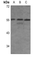 Western blot analysis of IRF3 (pS396) expression in HepG2 (A), mouse muscle (B), rat muscle (C) whole cell lysates.
Bioworld Biotech only provide peptides for our antibodies and do not provide additional peptide customization services.
Price/Size :
USD 368/1mg/vial

Tips:
For phospho antibody, we provide phospho peptide(0.5mg) and non-phospho peptide(0.5mg).Describe :
Blocking peptides are peptides that bind specifically to the target antibody and block antibody binding. These peptide usually contains the epitope recognized by the antibody. Antibodies bound to the blocking peptide no longer bind to the epitope on the target protein. This mechanism is useful when non-specific binding is an issue, for example, in Western blotting (WB) and Immunohistochemistry (IHC). By comparing the staining from the blocked antibody versus the antibody alone, one can see which staining is specific; Specific binding will be absent from the western blot or IHC performed with the neutralized antibody.Formula:
Synthetic peptide was lyophilized with 100% acetonitrile and is supplied as a powder. Reconstitute with 0.1 ml DI water for a final concentration of 10 mg/ml.The purity is >90%,tested by HPLC and MS.
Storage:
The freeze-dried powder is more stable. For short time at 2-8°C. For long term storage store at -20°C.
Note :
This product is for research use only (RUO only). Not for use in diagnostic or therapeutic procedures.