
 TGF beta 2 monoclonal antibody
  Datasheet
 COA
COA MSDS
MSDS SHIP
SHIP
Product Name :
TGF beta 2 monoclonal antibody Background :
Transforming growth factor-β (TGF-β) superfamily members are critical regulators of cell proliferation and differentiation, developmental patterning and morphogenesis, and disease pathogenesis . TGF-β elicits signaling through three cell surface receptors: type I (RI), type II (RII), and type III (RIII). Type I and type II receptors are serine/threonine kinases that form a heteromeric complex. In response to ligand binding, the type II receptors form a stable complex with the type I receptors allowing phosphorylation and activation of type I receptor kinases . The type III receptor, also known as betaglycan, is a transmembrane proteoglycan with a large extracellular domain that binds TGF-β with high affinity but lacks a cytoplasmic signaling domain . Expression of the type III receptor can regulate TGF-β signaling through presentation of the ligand to the signaling complex. The only known direct TGF-β signaling effectors are the Smad family proteins, which transduce signals from the cell surface directly to the nucleus to regulate target gene transcription .Three isoforms of TGF-β, designated TGF-β1, TGF-β2 and TGF-β3, are encoded by distinct genes and are expressed in a tissue specific manner . Each isoform is synthesized as a larger precursor protein containing a propeptide region that is removed prior to secretion. Mature TGF-β contains two polypeptides linked by disulfide bonds to form a protein of approximately 25 kDa. Product :
Mouse IgG1 kappa. Liquid in PBS, pH 7.3, 30% glycerol, and 0.01% sodium azide. Storage&Stability :
Store at 4°C short term. Aliquot and store at -20°C long term. Avoid freeze-thaw cycles. Specificity :
Recognizes endogenous levels of TGF beta 2 protein. Immunogen :
Recombinant fusion protein of human TGF beta 2. The exact sequence is proprietary. Conjugate :
Unconjugated Modification :
Unmodification
-
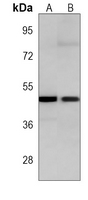 Western blot analysis of TGF beta 2 expression in 293 (A), K562 (B) whole cell lysates.
 -
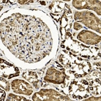 Immunohistochemical analysis of TGF beta 2 staining in human kidney formalin fixed paraffin embedded tissue section. The section was pre-treated using heat mediated antigen retrieval with sodium citrate buffer (pH 6.0). The section was then incubated with the antibody at room temperature and detected using an HRP conjugated compact polymer system. DAB was used as the chromogen. The section was then counterstained with haematoxylin and mounted with DPX.
Bioworld Biotech only provide peptides for our antibodies and do not provide additional peptide customization services.
Price/Size :
USD 368/1mg/vial

Tips:
For phospho antibody, we provide phospho peptide(0.5mg) and non-phospho peptide(0.5mg).Describe :
Blocking peptides are peptides that bind specifically to the target antibody and block antibody binding. These peptide usually contains the epitope recognized by the antibody. Antibodies bound to the blocking peptide no longer bind to the epitope on the target protein. This mechanism is useful when non-specific binding is an issue, for example, in Western blotting (WB) and Immunohistochemistry (IHC). By comparing the staining from the blocked antibody versus the antibody alone, one can see which staining is specific; Specific binding will be absent from the western blot or IHC performed with the neutralized antibody.Formula:
Synthetic peptide was lyophilized with 100% acetonitrile and is supplied as a powder. Reconstitute with 0.1 ml DI water for a final concentration of 10 mg/ml.The purity is >90%,tested by HPLC and MS.
Storage:
The freeze-dried powder is more stable. For short time at 2-8°C. For long term storage store at -20°C.
Note :
This product is for research use only (RUO only). Not for use in diagnostic or therapeutic procedures.