
 c-FOS monoclonal antibody
  Datasheet
 COA
COA MSDS
MSDS SHIP
SHIP
Product Name :
c-FOS monoclonal antibody Background :
The Fos family of nuclear oncogenes includes c-Fos, FosB, Fos-related antigen 1 (FRA1), and Fos-related antigen 2 (FRA2). While most Fos proteins exist as a single isoform, the FosB protein exists as two isoforms: full-length FosB and a shorter form, FosB2 (Delta FosB), which lacks the carboxy-terminal 101 amino acids. The expression of Fos proteins is rapidly and transiently induced by a variety of extracellular stimuli including growth factors, cytokines, neurotransmitters, polypeptide hormones, and stress. Fos proteins dimerize with Jun proteins (c-Jun, JunB, and JunD) to form Activator Protein-1 (AP-1), a transcription factor that binds to TRE/AP-1 elements and activates transcription. Fos and Jun proteins contain the leucine-zipper motif that mediates dimerization and an adjacent basic domain that binds to DNA. The various Fos/Jun heterodimers differ in their ability to transactivate AP-1 dependent genes. In addition to increased expression, phosphorylation of Fos proteins by Erk kinases in response to extracellular stimuli may further increase transcriptional activity. Phosphorylation of c-Fos at Ser32 and Thr232 by Erk5 increases protein stability and nuclear localization. Phosphorylation of FRA1 at Ser252 and Ser265 by Erk1/2 increases protein stability and leads to overexpression of FRA1 in cancer cells. Following growth factor stimulation, expression of FosB and c-Fos in quiescent fibroblasts is immediate, but very short-lived, with protein levels dissipating after several hours. FRA1 and FRA2 expression persists longer, and appreciable levels can be detected in asynchronously growing cells. Deregulated expression of c-Fos, FosB, or FRA2 can result in neoplastic cellular transformation; however, Delta FosB lacks the ability to transform cells. Product :
Liquid in 0.42% Potassium phosphate, 0.87% Sodium chloride, pH 7.3, 30% glycerol, and 0.01% sodium azide. Storage&Stability :
Store at 4°C short term. Aliquot and store at -20°C long term. Avoid freeze-thaw cycles. Specificity :
Recognizes endogenous levels of c-FOS protein. Immunogen :
Recombinant protein corresponding to human c-FOS. Conjugate :
Unconjugated Modification :
Unmodification
-
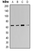 Western blot analysis of c-FOS expression in Hela (A), 293T (B), mouse brain (C), rat brain (D) whole cell lysates.
 -
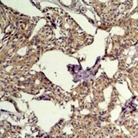 Immunohistochemical analysis of c-FOS staining in human breast cancer formalin fixed paraffin embedded tissue section. The section was pre-treated using heat mediated antigen retrieval with sodium citrate buffer (pH 6.0). The section was then incubated with the antibody at room temperature and detected using an HRP conjugated compact polymer system. DAB was used as the chromogen. The section was then counterstained with haematoxylin and mounted with DPX.
Bioworld Biotech only provide peptides for our antibodies and do not provide additional peptide customization services.
Price/Size :
USD 368/1mg/vial

Tips:
For phospho antibody, we provide phospho peptide(0.5mg) and non-phospho peptide(0.5mg).Describe :
Blocking peptides are peptides that bind specifically to the target antibody and block antibody binding. These peptide usually contains the epitope recognized by the antibody. Antibodies bound to the blocking peptide no longer bind to the epitope on the target protein. This mechanism is useful when non-specific binding is an issue, for example, in Western blotting (WB) and Immunohistochemistry (IHC). By comparing the staining from the blocked antibody versus the antibody alone, one can see which staining is specific; Specific binding will be absent from the western blot or IHC performed with the neutralized antibody.Formula:
Synthetic peptide was lyophilized with 100% acetonitrile and is supplied as a powder. Reconstitute with 0.1 ml DI water for a final concentration of 10 mg/ml.The purity is >90%,tested by HPLC and MS.
Storage:
The freeze-dried powder is more stable. For short time at 2-8°C. For long term storage store at -20°C.
Note :
This product is for research use only (RUO only). Not for use in diagnostic or therapeutic procedures.