
 PD-L1 polyclonal antibody
  Datasheet
 COA
COA MSDS
MSDS SHIP
SHIP
Product Name :
PD-L1 polyclonal antibody Background :
Engagement of CD28 by B7-1 (CD80) or B7-2 (CD86) in the presence of antigen promotes T cell proliferation, cytokine production, differentiation of effector T cells, and the induction of Bcl-x, a promoter of T cell survival. Conversely, engagement of CTLA4 by B7-1 or B7-2 may inhibit proliferation and IL-2 production. Pdcd-1L1 (programmed cell death ligand-1), also known as B7-H1 or PD-L1, is 290 amino acid type I transmembrane protein which is 20% and 15% identical to B7-1 and B7-2, respectively. Pdcd-1L2 has immunoglobulin V-like and C-like domains and a 30 amino acid cytoplasmic tail. It does not bind CD28, cytotoxic T-lymphocyte A4 or ICOS (inducible co-stimulator). IL-2, although produced in small amounts, is required for the effect of Pdcd-1L1 co-stimulation. The gene which encodes Pdcd-1L1 maps to human chromosome 9p24. Pdcd-1L2 (programmed cell death ligand-2) is a 73 amino acid protein which contains a signal sequence, IgV- and IgC-like domains, a transmembrane region and a cytoplasmic region. The gene which encodes Pdcd-1L2 maps to human chromosome 9p24.2. The constitutive expression of Pdcd-1L1 and Pdcd-1L2 on paren-chymal cells of heart, lung and kidney suggests that the Pdcd-1-Pdcd-L system could provide unique negative signaling to help prevent autoimmune disease. Product :
Rabbit IgG, 1mg/ml in PBS with 0.02% sodium azide, 50% glycerol, pH7.2 Storage&Stability :
Store at 4°C after thawing. Aliquot store at -20°C or -80°C. Avoid repeated freeze / thaw cycles. Specificity :
PD-L1 polyclonal antibody detects endogenous levels of PD-L1 protein. Immunogen :
recombinant protein Conjugate :
Unconjugated Modification :
Unmodification
-
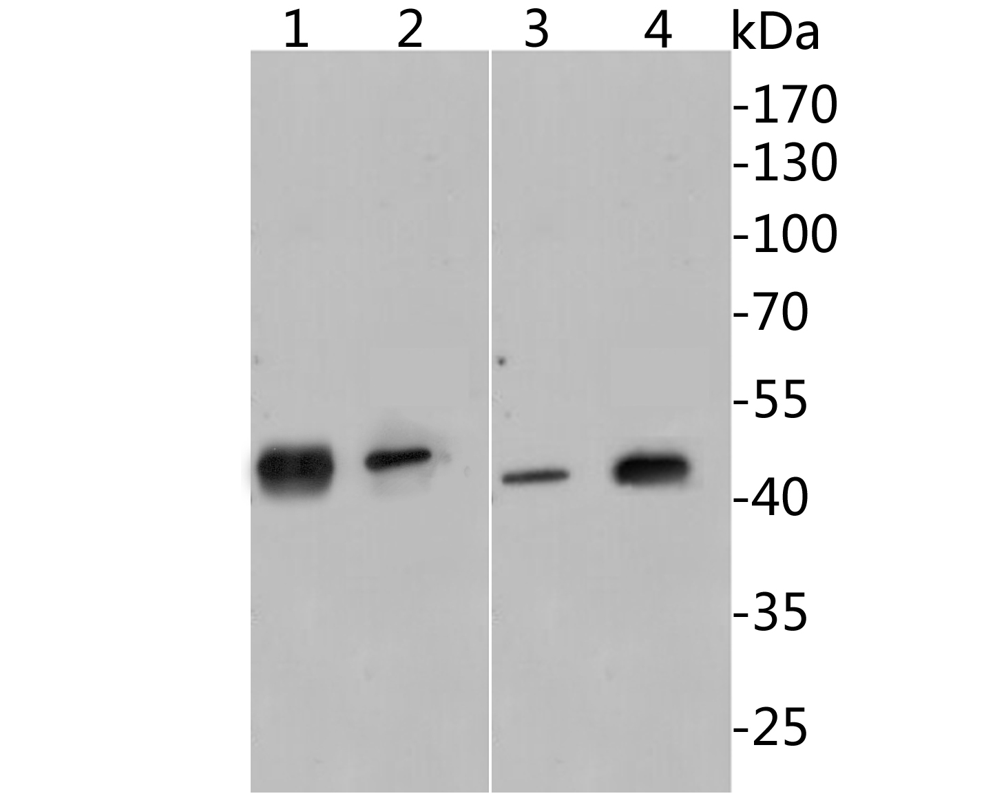 Western blot analysis of PD-L1 on different lysates. Proteins were transferred to a PVDF membrane and blocked with 5% BSA in PBS for 1 hour at room temperature. The primary antibody( 1:500) was used in 5% BSA at room temperature for 2 hours. Lane 1: A549 cell lysates(40ug) Lane 2: MCF-7 cell lysates(40ug) Lane 3: Mouse placenta tissue lysates(40ug) Lane 4: Mouse lung tissue lysates
 -
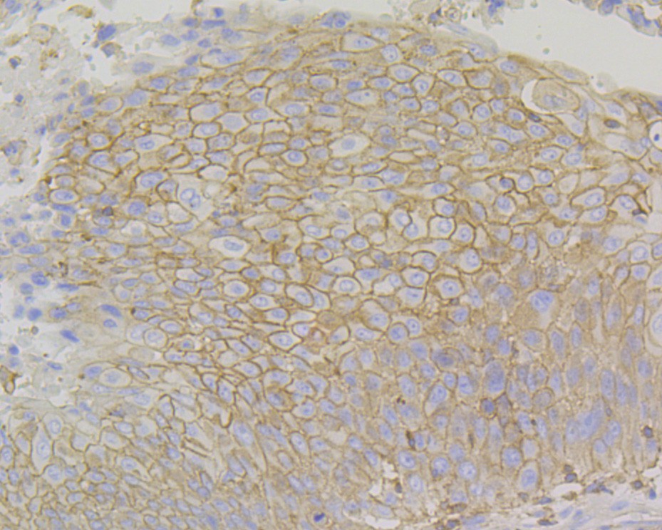 Immunohistochemical analysis of paraffin-embedded human non-small cell lung cancer tissue using anti-PD-L1 antibody. The section was pre-treated using heat mediated antigen retrieval with Tris-EDTA buffer (pH 8.0-8.4) for 20 minutes.The tissues were blocked in 5% BSA for 30 minutes at room temperature, washed with ddH2O and PBS, and then probed with the primary antibody (1:200) for 30 minutes at room temperature. The detection was performed using an HRP conjugated compact polymer system. DAB was used as the chromogen. Tissues were counterstained with hematoxylin and mounted with DPX.
Bioworld Biotech only provide peptides for our antibodies and do not provide additional peptide customization services.
Price/Size :
USD 368/1mg/vial

Tips:
For phospho antibody, we provide phospho peptide(0.5mg) and non-phospho peptide(0.5mg).Describe :
Blocking peptides are peptides that bind specifically to the target antibody and block antibody binding. These peptide usually contains the epitope recognized by the antibody. Antibodies bound to the blocking peptide no longer bind to the epitope on the target protein. This mechanism is useful when non-specific binding is an issue, for example, in Western blotting (WB) and Immunohistochemistry (IHC). By comparing the staining from the blocked antibody versus the antibody alone, one can see which staining is specific; Specific binding will be absent from the western blot or IHC performed with the neutralized antibody.Formula:
Synthetic peptide was lyophilized with 100% acetonitrile and is supplied as a powder. Reconstitute with 0.1 ml DI water for a final concentration of 10 mg/ml.The purity is >90%,tested by HPLC and MS.
Storage:
The freeze-dried powder is more stable. For short time at 2-8°C. For long term storage store at -20°C.
Note :
This product is for research use only (RUO only). Not for use in diagnostic or therapeutic procedures.