
 PD1 monoclonal antibody
  Datasheet
 COA
COA MSDS
MSDS SHIP
SHIP
Product Name :
PD1 monoclonal antibody Background :
Pdcd-1 (Programmed Cell Death-1 protein), also designated CD279, is a type I transmembrane receptor and a member of the immunoglobin gene superfamily. Pdcd-1 contains an immunoreceptor tyrosine-based inhibitory motif (ITIM) within the cytoplasmic domain, which is conserved between the mouse and human homologs. Expression of Pdcd-1 is detected in mouse thymus, and it is induced in stimulated B and T cell lines, where it may play a role in the negative regulation of various immune responses. Receptors such as Pdcd-1 function by recruiting tyrosine phosphatases, including SHP-1 and SHIP, which are responsible for altering various B cell responses. Additionally, in activated lymphocytes, Pdcd-1 mediates the activation of the classical type of programmed cell death. Product :
Mouse IgG1, 1mg/ml in PBS with 0.02% sodium azide, 50% glycerol, pH7.2 Storage&Stability :
Store at +4°C after thawing. Aliquot store at -20°C or -80°C. Avoid repeated freeze / thaw cycles. Specificity :
PD1 monoclonal antibody detects endogenous levels of PD1 protein. Immunogen :
Recombinant protein Conjugate :
Unconjugated Modification :
Unmodification
-
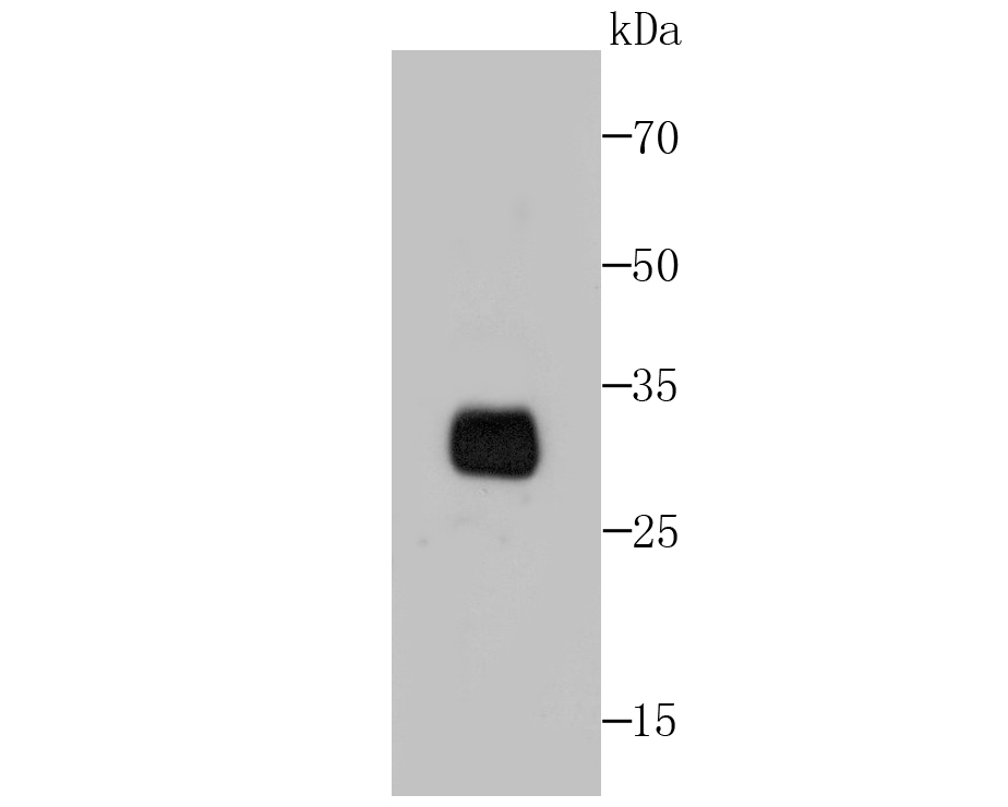 Western blot analysis of PD1 on PD1 transfected 293FT cell lysate using anti-PD1 antibody at 1/1,000 dilution.
 -
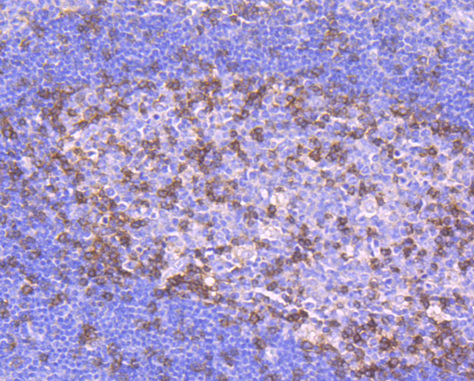 Immunohistochemical analysis of paraffin-embedded human tonsil tissue using anti-PD1 antibody. Counter stained with hematoxylin.
The impact of tobacco exposure on tumor microenvironment and prognosis in lung adenocarcinoma by integrative analysis of multi-omics data
PMCID: Pubmed No.:34700112
Programmed cell death‑1/programmed cell death‑ligand 1 inhibitors exert antiapoptosis and antiinflammatory activity in lipopolysaccharide stimulated murine alveolar macrophages
PMCID: Pubmed No.:33680122
Bioworld Biotech only provide peptides for our antibodies and do not provide additional peptide customization services.
Price/Size :
USD 368/1mg/vial

Tips:
For phospho antibody, we provide phospho peptide(0.5mg) and non-phospho peptide(0.5mg).Describe :
Blocking peptides are peptides that bind specifically to the target antibody and block antibody binding. These peptide usually contains the epitope recognized by the antibody. Antibodies bound to the blocking peptide no longer bind to the epitope on the target protein. This mechanism is useful when non-specific binding is an issue, for example, in Western blotting (WB) and Immunohistochemistry (IHC). By comparing the staining from the blocked antibody versus the antibody alone, one can see which staining is specific; Specific binding will be absent from the western blot or IHC performed with the neutralized antibody.Formula:
Synthetic peptide was lyophilized with 100% acetonitrile and is supplied as a powder. Reconstitute with 0.1 ml DI water for a final concentration of 10 mg/ml.The purity is >90%,tested by HPLC and MS.
Storage:
The freeze-dried powder is more stable. For short time at 2-8°C. For long term storage store at -20°C.
Note :
This product is for research use only (RUO only). Not for use in diagnostic or therapeutic procedures.