
 14-3-3 ζ (V52) polyclonal antibody
  Datasheet
 COA
COA MSDS
MSDS SHIP
SHIP
Product Name :
14-3-3 ζ (V52) polyclonal antibody Background :
14-3-3 proteins regulate many cellular processes relevant to cancer biology, notably apoptosis, mitogenic signaling and cell-cycle checkpoints. Seven isoforms comprise this family of signaling intermediates, denoted 14-3-3 β, γ, ε, ζ, η, θ and σ. 14-3-3 proteins form dimers that present two binding sites for ligand proteins, thereby bringing together two proteins that may not otherwise associate. These ligands largely share a 14-3-3 consensus binding motif and exhibit serine/threonine phosphorylation. 14-3-3 proteins function in broad regulation of these ligand proteins, by cytoplasmic sequestration, occupation of interaction domains and import/export sequences, prevention of degradation, activation/repression of enzymatic activity and facilitation of protein modification, and thus loss of expression contributes to a vast array of pathogenic cellular activities. Product :
Rabbit IgG, 1mg/ml in PBS with 0.02% sodium azide, 50% glycerol, pH7.2 Storage&Stability :
Store at 4°C short term. Aliquot and store at -20°C long term. Avoid freeze-thaw cycles. Specificity :
14-3-3 ζ (V52) polyclonal antibody detects endogenous levels of 14-3-3 protein zeta/delta. Immunogen :
Synthetic peptide, corresponding to amino acids 20-70 of Human 14-3-3 ζ. Conjugate :
Unconjugated Modification :
Unmodification
-
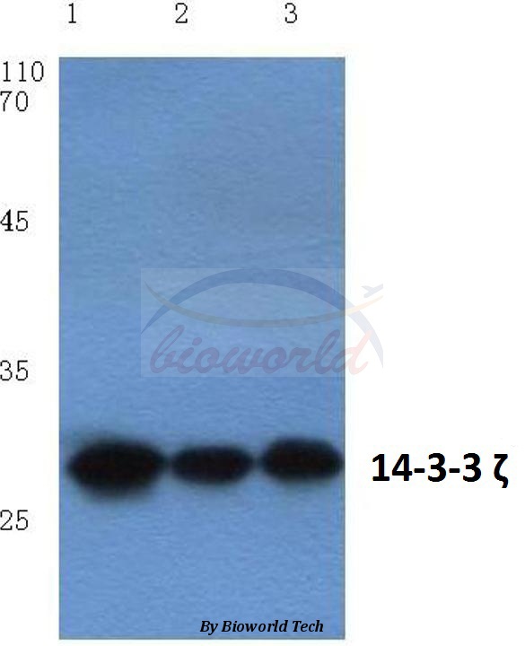 Western blot (WB) analysis of 14-3-3 ζ (V52) polyclonal antibody at 1:500 dilution Lane1:Hela cell lysate Lane2:Mouse brain tissue lysate Lane3:Rat kidney tissue lysate
 -
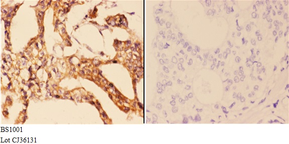 Immunohistochemistry (IHC) analyzes of 14-3-3 ζ (V52) pAb in paraffin-embedded human breast carcinoma tissue at 1:50,showing cytoplasmic and nuclear staining.Negative control (the right)Using PBS instead of primary antibody, secondary antibody is Goat Anti-Rabbit IgG-biotin followed by avidin-peroxidase.
 -
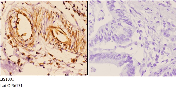 Immunohistochemistry (IHC) analyzes of 14-3-3 ζ (V52) pAb in paraffin-embedded human breast carcinoma tissue at 1:50,showing cytoplasmic and nuclear staining.Negative control (the right)Using PBS instead of primary antibody, secondary antibody is Goat Anti-Rabbit IgG-biotin followed by avidin-peroxidase.
 -
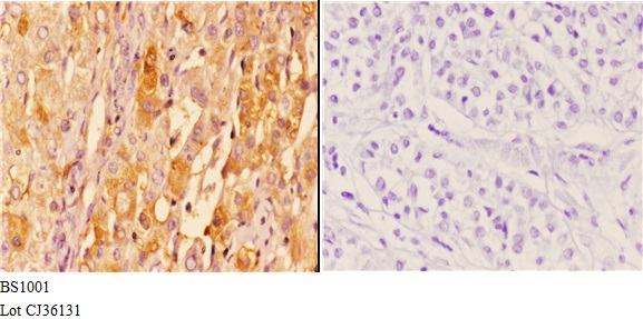 Immunohistochemistry (IHC) analyzes of 14-3-3 ζ (V52) pAb in paraffin-embedded human breast carcinoma tissue at 1:50,showing cytoplasmic and nuclear staining.Negative control (the right)Using PBS instead of primary antibody, secondary antibody is Goat Anti-Rabbit IgG-biotin followed by avidin-peroxidase.
Plasma Membrane H+-ATPase and 14-3-3 Isoforms of Arabidopsis Leaves: Evidence for Isoform Specificity in the 14-3-3/H+ -ATPase Interaction
PMCID: Pubmed No.:15509843
Identification of potential pathways involved in the induction of cell cycle arrest and apoptosis by a new 4-arylidene curcumin analogue T63 in lung cancer cells: a comparative proteomic analysis.
PMCID: Pubmed No.:24651282
Proteomic analysis on infantile spasm and prenatal stress
PMCID: Pubmed No.:23028951
SET-mediated NDRG1 inhibition is involved in acquisition of epithelial-to-mesenchymal transition phenotype and cisplatin resistance in human lung cancer cell
PMCID: Pubmed No.:25152373
SET-mediated NDRG1 inhibition is involved in acquisition of epithelial-to-mesenchymal transition phenotype and cisplatin resistance in human lung cancer cell
PMCID: Pubmed No.:25152373
Identification of potential pathways involved in the induction of cell cycle arrest and apoptosis by a new 4-arylidene curcumin analogue T63 in lung cancer cells: a comparative proteomic analysis.
PMCID: Pubmed No.:24651282
Bioworld Biotech only provide peptides for our antibodies and do not provide additional peptide customization services.
Price/Size :
USD 368/1mg/vial

Tips:
For phospho antibody, we provide phospho peptide(0.5mg) and non-phospho peptide(0.5mg).Describe :
Blocking peptides are peptides that bind specifically to the target antibody and block antibody binding. These peptide usually contains the epitope recognized by the antibody. Antibodies bound to the blocking peptide no longer bind to the epitope on the target protein. This mechanism is useful when non-specific binding is an issue, for example, in Western blotting (WB) and Immunohistochemistry (IHC). By comparing the staining from the blocked antibody versus the antibody alone, one can see which staining is specific; Specific binding will be absent from the western blot or IHC performed with the neutralized antibody.Formula:
Synthetic peptide was lyophilized with 100% acetonitrile and is supplied as a powder. Reconstitute with 0.1 ml DI water for a final concentration of 10 mg/ml.The purity is >90%,tested by HPLC and MS.
Storage:
The freeze-dried powder is more stable. For short time at 2-8°C. For long term storage store at -20°C.
Note :
This product is for research use only (RUO only). Not for use in diagnostic or therapeutic procedures.