
 Bak (A2) polyclonal antibody
  Datasheet
 COA
COA MSDS
MSDS SHIP
SHIP
Product Name :
Bak (A2) polyclonal antibody Background :
Bak or Bcl2 homologous antagonist is a member of the Bcl2 family of proteins. The Bcl-2 related proteins interact with one another through the formation of homo and heterodimers. The susceptibility of cells to apoptotic stimuli is thought to be controlled by the relative ratios of the different Bcl2 family proteins. Bak has been demonstrated to accelerate the rate of apoptosis in growth factor deprived murine lymphoid, neuronal and fibroblastic cell lines. This protein localizes to mitochondria, and functions to induce apoptosis. It interacts with and accelerates the opening of the mitochondrial voltage-dependent anion channel, which leads to a loss in membrane potential and the release of cytochrome c. Product :
Rabbit IgG, 1mg/ml in PBS with 0.02% sodium azide, 50% glycerol, pH7.2 Storage&Stability :
Store at 4°C short term. Aliquot and store at -20°C long term. Avoid freeze-thaw cycles. Specificity :
Bak (A2) polyclonal antibody detects endogenous levels of Bak protein. Immunogen :
Synthetic peptide, corresponding to the N-terminal of Human Bak. Conjugate :
Unconjugated Modification :
Unmodification
-
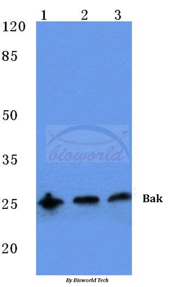 Western blot (WB) analysis of Bak (A2) polyclonal antibody at 1:500 dilution Lane1:Hela cell lysate Lane2:NIH-3T3 cell lysate Lane3:Rat kidney tissue lysate
 -
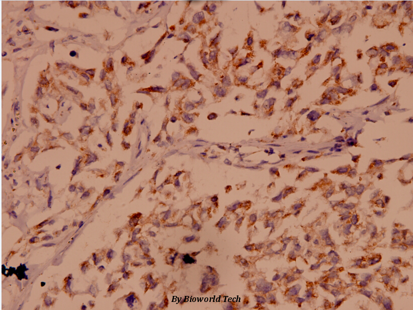 Immunohistochemistry (IHC) analyzes of Bak (A2) pAb in paraffin-embedded human breast carcinoma tissue at 1:100.
Methylated actinomycin D, a novel actinomycin D analog induces apoptosis in HepG2 cells through fas- and mitochondria-mediated pathways†
PMCID: Pubmed No.:22821714
Silencing Prion Protein in MDA-MB-435 Breast Cancer Cells Leads to Pleiotropic Cellular Responses to Cytotoxic Stimuli
PMCID: Pubmed No.:23133614
Tetrandrine Induces Mitochondria-Mediated Apoptosis in Human Gastric Cancer BGC-823 Cells
PMCID: Pubmed No.:24098511
Benzo(a)pyrene-7,8-diol-9,10-epoxide induced p53-independent necrosis via the mitochondria-associated pathway involving Bax and Bak activation.
PMCID: Pubmed No.:24837741
Role of Bax/Bcl-2 family members in green tea polyphenol induced necroptosis of p53-deficient Hep3B cells
PMCID: Pubmed No.:24839007
Tetrandrine Induces Mitochondria-Mediated Apoptosis in Human Gastric Cancer BGC-823 Cells
PMCID: Pubmed No.:24098511
HDAC10 promotes lung cancer proliferation via AKT phosphorylation
PMCID: Pubmed No.:27449083
Proteomic analysis reveals ginsenoside Rb1 attenuates myocardial ischemia/reperfusion injury through inhibiting ROS production from mitochondrial complex I.
PMCID: Pubmed No.:33408776
Nuclear respiratory factor 1 protects H9C2 cells against hypoxia-induced apoptosis via the death receptor pathway and mitochondrial pathway
PMCID: Pubmed No.:33913583
Combination of oxytetracycline and quinocetone synergistically induces hepatotoxicity via generation of reactive oxygen species and activation of mitochondrial pathway
PMCID: Pubmed No.:34348565
Bioworld Biotech only provide peptides for our antibodies and do not provide additional peptide customization services.
Price/Size :
USD 368/1mg/vial

Tips:
For phospho antibody, we provide phospho peptide(0.5mg) and non-phospho peptide(0.5mg).Describe :
Blocking peptides are peptides that bind specifically to the target antibody and block antibody binding. These peptide usually contains the epitope recognized by the antibody. Antibodies bound to the blocking peptide no longer bind to the epitope on the target protein. This mechanism is useful when non-specific binding is an issue, for example, in Western blotting (WB) and Immunohistochemistry (IHC). By comparing the staining from the blocked antibody versus the antibody alone, one can see which staining is specific; Specific binding will be absent from the western blot or IHC performed with the neutralized antibody.Formula:
Synthetic peptide was lyophilized with 100% acetonitrile and is supplied as a powder. Reconstitute with 0.1 ml DI water for a final concentration of 10 mg/ml.The purity is >90%,tested by HPLC and MS.
Storage:
The freeze-dried powder is more stable. For short time at 2-8°C. For long term storage store at -20°C.
Note :
This product is for research use only (RUO only). Not for use in diagnostic or therapeutic procedures.