
 AIF-M1 (Y85) polyclonal antibody
  Datasheet
 COA
COA MSDS
MSDS SHIP
SHIP
Product Name :
AIF-M1 (Y85) polyclonal antibody Background :
Apoptosis is characterized by several morphological nuclear changes including chromatin condensation and nuclear fragmentation. These changes are triggered by the activation of members of caspase family, caspase activated DNase, and several novel proteins. A novel gene, the product of which causes chromatin condensation and DNA fragmentation, was recently identified, cloned, and designated apoptosis inducing factor (AIF). Like the critical molecules, cytochrome c and caspase 9, in apoptosis, AIF localizes in mitochondria. AIF translocates to the nucleus when apoptosis is induced and induces mitochondria to release the apoptogenic proteins cytochrome c and caspase 9. AIF induces chromatin condensation and large scale DNA fragmentation, which are the hallmarks of apoptosis, of the isolated nucleus and the nucleus in live cells by microinjection and apoptosis stimuli. AIF is highly conserved between human and mouse and widely expressed. Product :
Rabbit IgG, 1mg/ml in PBS with 0.02% sodium azide, 50% glycerol, pH7.2 Storage&Stability :
Store at 4°C short term. Aliquot and store at -20°C long term. Avoid freeze-thaw cycles. Specificity :
AIF-M1 (Y85) polyclonal antibody detects endogenous levels of AIF-M1 protein. Immunogen :
Synthetic peptide, corresponding to amino acids 61-110 of Human AIF-M1. Conjugate :
Unconjugated Modification :
Unmodification
-
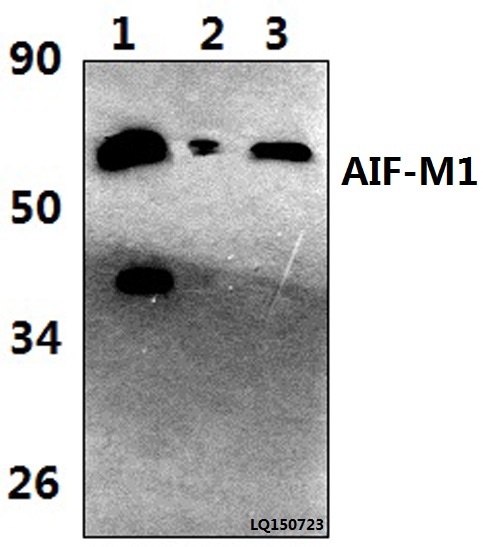 Western blot (WB) analysis of AIF-M1 (Y85) polyclonal antibody at 1:500 dilution Lane1:The liver tissue lysate of Mouse(39ug) Lane2:MCF-7 whole cell lysate(40ug) Lane3:H9C2 whole cell lysate(40ug)
 -
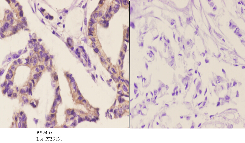 Immunohistochemistry (IHC) analyzes of AIF-M1 (Y85) pAb in paraffin-embedded human breast carcinoma tissue at 1:50.showing cytoplasmic and nucleus staining. Negative control (the right)Using PBS instead of primary antibody, secondary antibody is Goat Anti-Rabbit IgG-biotin followed by avidin-peroxidase.
 -
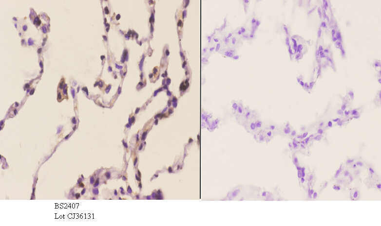 Immunohistochemistry (IHC) analyzes of AIF-M1 (Y85) pAb in paraffin-embedded human breast carcinoma tissue at 1:50.showing cytoplasmic and nucleus staining. Negative control (the right)Using PBS instead of primary antibody, secondary antibody is Goat Anti-Rabbit IgG-biotin followed by avidin-peroxidase.
 -
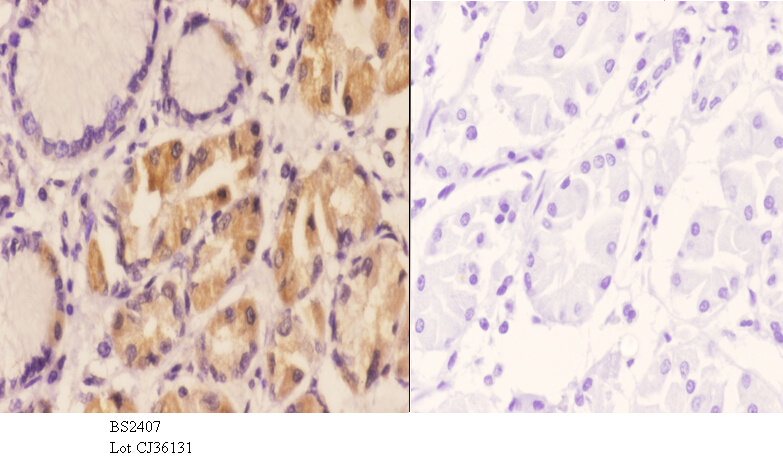 Immunohistochemistry (IHC) analyzes of AIF-M1 (Y85) pAb in paraffin-embedded human breast carcinoma tissue at 1:50.showing cytoplasmic and nucleus staining. Negative control (the right)Using PBS instead of primary antibody, secondary antibody is Goat Anti-Rabbit IgG-biotin followed by avidin-peroxidase.
Bioworld Biotech only provide peptides for our antibodies and do not provide additional peptide customization services.
Price/Size :
USD 368/1mg/vial

Tips:
For phospho antibody, we provide phospho peptide(0.5mg) and non-phospho peptide(0.5mg).Describe :
Blocking peptides are peptides that bind specifically to the target antibody and block antibody binding. These peptide usually contains the epitope recognized by the antibody. Antibodies bound to the blocking peptide no longer bind to the epitope on the target protein. This mechanism is useful when non-specific binding is an issue, for example, in Western blotting (WB) and Immunohistochemistry (IHC). By comparing the staining from the blocked antibody versus the antibody alone, one can see which staining is specific; Specific binding will be absent from the western blot or IHC performed with the neutralized antibody.Formula:
Synthetic peptide was lyophilized with 100% acetonitrile and is supplied as a powder. Reconstitute with 0.1 ml DI water for a final concentration of 10 mg/ml.The purity is >90%,tested by HPLC and MS.
Storage:
The freeze-dried powder is more stable. For short time at 2-8°C. For long term storage store at -20°C.
Note :
This product is for research use only (RUO only). Not for use in diagnostic or therapeutic procedures.