
 PI3K p110α (R519) polyclonal antibody
  Datasheet
 COA
COA MSDS
MSDS SHIP
SHIP
Product Name :
PI3K p110α (R519) polyclonal antibody Background :
Phosphatidylinositol 3-kinase (PI 3-kinase) is composed of 85 kDa (p85) and 110 kDa (p110) subunits. p85 lacks PI 3-kinase activity and acts as an adapter, coupling p110 to activated protein tyrosine kinase. Two forms of p85 have been described (p85α and p85β), each possessing one SH3 and two SH2 domains. Various p110 isoforms have been identified. p110α and p110β interact with p85α, and p110α has also been shown to interact with p85β in vitro. p110δ expression is restricted to white blood cells. It has been shown to bind p85α and β, but it apparently does not phosphorylate these subunits. p110δ seems to have the capacity to autophosphorylate. p110γ does not interact with the p85 subunits. It has been shown to be activated by α and βγ heterotrimeric G proteins._x000D_ Product :
Rabbit IgG, 1mg/ml in PBS with 0.02% sodium azide, 50% glycerol, pH7.2 Storage&Stability :
Store at 4°C short term. Aliquot and store at -20°C long term. Avoid freeze-thaw cycles. Specificity :
PI 3-kinase p110α (R519) polyclonal antibody detects endogenous levels of PI 3-kinase p110α protein. Immunogen :
Synthetic peptide, corresponding to amino acids 500-550 of Human PI3K p110α. Conjugate :
Unconjugated Modification :
Unmodification
-

-
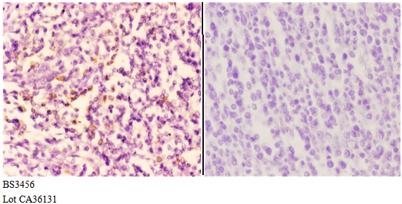 Immunohistochemistry (IHC) analyzes of PI3K p110α (R519) pAb in paraffin-embedded human tonsil carcinoma tissue at 1:50,showing cytoplasmic and nuclear staining.Negative control (the right)Using PBS instead of primary antibody, secondary antibody is Goat Anti-Rabbit IgG-biotin followed by avidin-peroxidase.
 -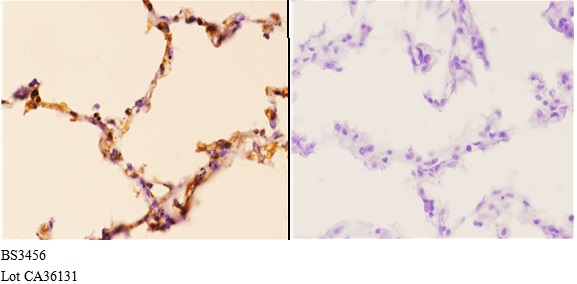
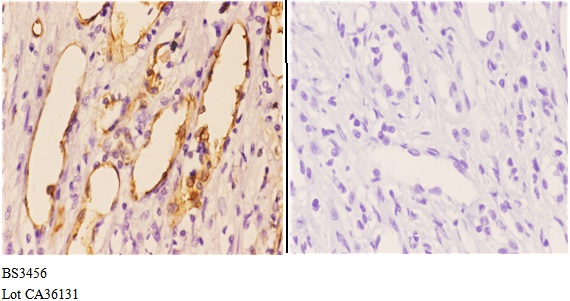 Immunohistochemistry (IHC) analyzes of PI3K p110α (R519) pAb in paraffin-embedded human tonsil carcinoma tissue at 1:50,showing cytoplasmic and nuclear staining.Negative control (the right)Using PBS instead of primary antibody, secondary antibody is Goat Anti-Rabbit IgG-biotin followed by avidin-peroxidase.
Estrogen-related receptor (ERR) γ protects against puromycin aminonucleoside-induced podocyte apoptosis by targeting PI3K/Akt signaling
PMCID: Pubmed No.:27417234
DIFFERENTIATED EMBRYONIC CHONDROCYTE EXPRESSED GENE-1 IS A CENTRAL SIGNALING COMPONENT IN THE DEVELOPMENT OF COLLAGEN-INDUCED RHEUMATOID ARTHRITIS
PMCID: Pubmed No.:36739947
Bioworld Biotech only provide peptides for our antibodies and do not provide additional peptide customization services.
Price/Size :
USD 368/1mg/vial

Tips:
For phospho antibody, we provide phospho peptide(0.5mg) and non-phospho peptide(0.5mg).Describe :
Blocking peptides are peptides that bind specifically to the target antibody and block antibody binding. These peptide usually contains the epitope recognized by the antibody. Antibodies bound to the blocking peptide no longer bind to the epitope on the target protein. This mechanism is useful when non-specific binding is an issue, for example, in Western blotting (WB) and Immunohistochemistry (IHC). By comparing the staining from the blocked antibody versus the antibody alone, one can see which staining is specific; Specific binding will be absent from the western blot or IHC performed with the neutralized antibody.Formula:
Synthetic peptide was lyophilized with 100% acetonitrile and is supplied as a powder. Reconstitute with 0.1 ml DI water for a final concentration of 10 mg/ml.The purity is >90%,tested by HPLC and MS.
Storage:
The freeze-dried powder is more stable. For short time at 2-8°C. For long term storage store at -20°C.
Note :
This product is for research use only (RUO only). Not for use in diagnostic or therapeutic procedures.