
 ATP6V1G3 polyclonal antibody
  Datasheet
 COA
COA MSDS
MSDS SHIP
SHIP
Product Name :
ATP6V1G3 polyclonal antibody Background :
Vacuolar-type H+-ATPase (V-ATPase) is a multisubunit enzyme responsible for acidification of eukaryotic intracellular organelles. V-ATPases pump protons against an electrochemical gradient, while F-ATPases reverse the process, thereby synthesizing ATP. A peripheral V1 domain, which is responsible for ATP hydrolysis, and a integral V0 domain, which is responsible for proton translocation, compose V-ATPase. Nine subunits (A–H) make up the V1 domain and five subunits (a, d, c, c' and c") make up the V0 domain. Like F-ATPase, V-ATPase most likely operates through a rotary mechanism. In yeast, the V-ATPase G subunit is a soluble subunit that shares homology with the F-ATPase G subunit and may be part of a connection stalk between V1 and V0. The G2 isoform of the G subunit associates with the pore-forming a1C-subunit of L-type calcium channel and aids in proper membrane targeting of the calcium channel. The genes encoding the G1 and G2 V-ATPase subunits map to chromosomes 9q33.1 and 6p21.3, respectively. Product :
Rabbit IgG, 1mg/ml in PBS with 0.02% sodium azide, 50% glycerol, pH7.2 Storage&Stability :
Store at 4°C short term. Aliquot and store at -20°C long term. Avoid freeze-thaw cycles. Specificity :
ATP6V1G3 polyclonal antibody detects endogenous levels of ATP6V1G3 protein. Immunogen :
A synthetic peptide corresponding to residues in Human ATP6V1G3. Conjugate :
Unconjugated Modification :
Unmodification
-
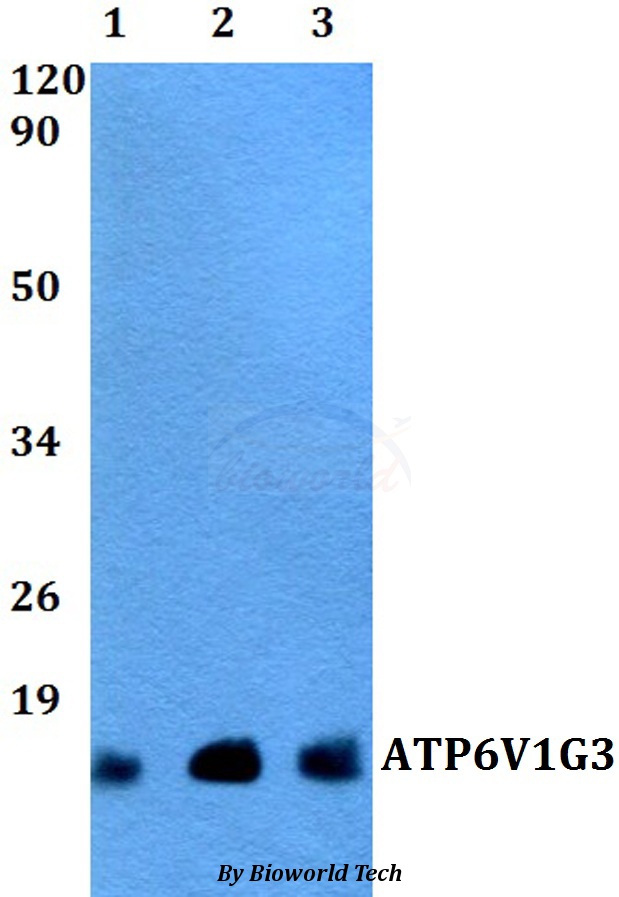 Western blot (WB) analysis of ATP6V1G3 polyclonal antibody at 1:500 dilution Lane1:HEK293T whole cell lysate Lane2:NIH-3T3 whole cell lysate Lane3:PC12 whole cell lysate
Bioworld Biotech only provide peptides for our antibodies and do not provide additional peptide customization services.
Price/Size :
USD 368/1mg/vial

Tips:
For phospho antibody, we provide phospho peptide(0.5mg) and non-phospho peptide(0.5mg).Describe :
Blocking peptides are peptides that bind specifically to the target antibody and block antibody binding. These peptide usually contains the epitope recognized by the antibody. Antibodies bound to the blocking peptide no longer bind to the epitope on the target protein. This mechanism is useful when non-specific binding is an issue, for example, in Western blotting (WB) and Immunohistochemistry (IHC). By comparing the staining from the blocked antibody versus the antibody alone, one can see which staining is specific; Specific binding will be absent from the western blot or IHC performed with the neutralized antibody.Formula:
Synthetic peptide was lyophilized with 100% acetonitrile and is supplied as a powder. Reconstitute with 0.1 ml DI water for a final concentration of 10 mg/ml.The purity is >90%,tested by HPLC and MS.
Storage:
The freeze-dried powder is more stable. For short time at 2-8°C. For long term storage store at -20°C.
Note :
This product is for research use only (RUO only). Not for use in diagnostic or therapeutic procedures.