
 ATM (phospho-S1987) polyclonal antibody
  Datasheet
 COA
COA MSDS
MSDS SHIP
SHIP
Product Name :
ATM (phospho-S1987) polyclonal antibody Background :
The phosphatidylinositol kinase (PIK) family members fall into two distinct subgroups. The first subgroup contains proteins such as the PI 3- and PI 4-kinases and the second group comprises the PIK-related kinases. The PIK-related kinases include Atm, DNA-PKCS and FRAP. These proteins have in common a region of homology at their carboxy-termini that is not present in the PI 3- and PI 4-kinases. The Atm gene is mutated in the autosomal recessive disorder ataxia telangiectasia (AT) that is characterized by cerebellar degeneration (ataxia) and the appearance of dilated blood vessels (telangiec-tases) in the conjunctivae of the eyes. AT cells are hypersensitive to ionizing radiation, impaired in mediating the inhibition of DNA synthesis and display delays in p53 induction. Product :
Rabbit IgG, 1mg/ml in PBS with 0.02% sodium azide, 50% glycerol, pH7.2 Storage&Stability :
Store at 4°C short term. Aliquot and store at -20°C long term. Avoid freeze-thaw cycles. Specificity :
p-ATM (S1987) polyclonal antibody detects endogenous levels of ATM protein only when phosphorylated at ser1987. Immunogen :
A synthetic peptide corresponding to residues in Human ATM around the phosphorylation site of ser1987. Conjugate :
Unconjugated Modification :
Phosphorylation
-
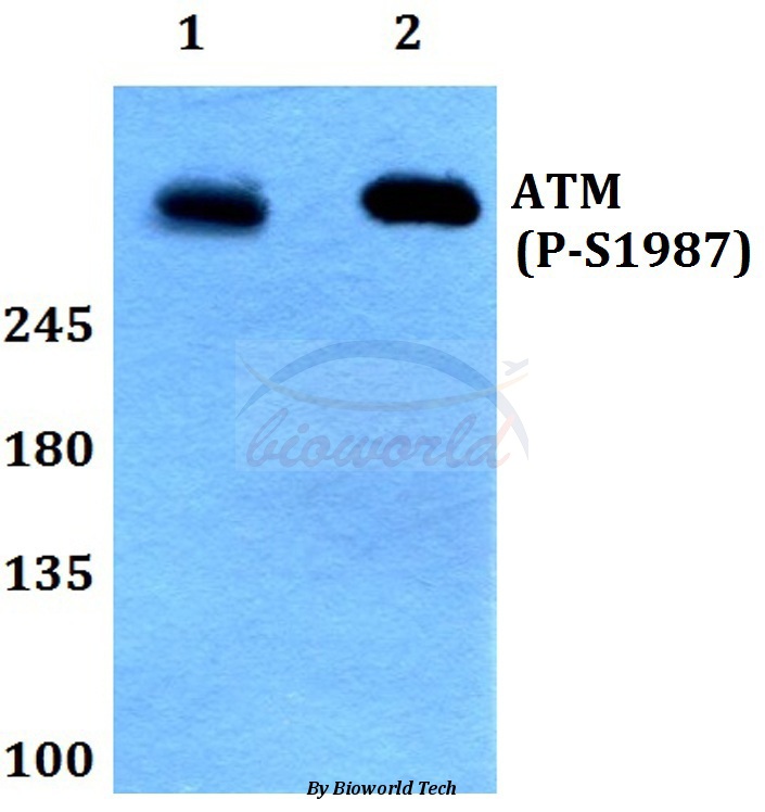 Western blot (WB) analysis of p-ATM (S1987) polyclonal antibody at 1:500 dilution Lane1:HEK293T whole cell lysate treated with UV(24h) Lane2:Raw264.7 whole cell lysate treated with UV(24h)
 -
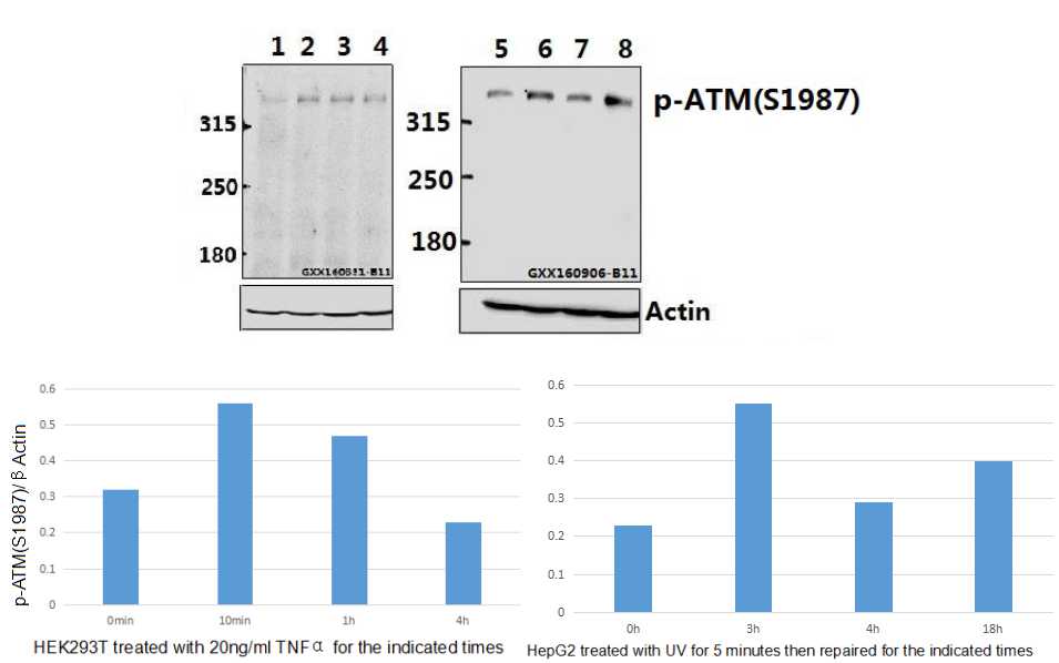 Western blot (WB) analysis of ATM (phospho-S1987) pAb at 1:500 dilution Lane1:HEK293T whole cell lysate(40ug) Lane2:HEK293T treated with TNFα(20ng/ml) for 10 minutes whole cell lysate Lane3:HEK293T treated with TNFα(20ng/ml) for 1 hour whole cell lysate Lane4:HEK293T treated with TNFα(20ng/ml) for 4 hours whole cell lysate Lane5:HepG2 whole cell lysate(40ug) Lane6:HepG2 treated with UV for 5 minutes then repaired for 3 hours whole cell lysate(40ug) Lane7:HepG2 treated with UV for 5 minutes then repaired for 4 hours whole cell lysate(40ug) Lane8:HepG2 treated with UV for 5 minutes then repaired for 18 hours whole cell lysate(40ug)
Bioworld Biotech only provide peptides for our antibodies and do not provide additional peptide customization services.
Price/Size :
USD 368/1mg/vial

Tips:
For phospho antibody, we provide phospho peptide(0.5mg) and non-phospho peptide(0.5mg).Describe :
Blocking peptides are peptides that bind specifically to the target antibody and block antibody binding. These peptide usually contains the epitope recognized by the antibody. Antibodies bound to the blocking peptide no longer bind to the epitope on the target protein. This mechanism is useful when non-specific binding is an issue, for example, in Western blotting (WB) and Immunohistochemistry (IHC). By comparing the staining from the blocked antibody versus the antibody alone, one can see which staining is specific; Specific binding will be absent from the western blot or IHC performed with the neutralized antibody.Formula:
Synthetic peptide was lyophilized with 100% acetonitrile and is supplied as a powder. Reconstitute with 0.1 ml DI water for a final concentration of 10 mg/ml.The purity is >90%,tested by HPLC and MS.
Storage:
The freeze-dried powder is more stable. For short time at 2-8°C. For long term storage store at -20°C.
Note :
This product is for research use only (RUO only). Not for use in diagnostic or therapeutic procedures.