
 MYL3 polyclonal antibody
  Datasheet
 COA
COA MSDS
MSDS SHIP
SHIP
Product Name :
MYL3 polyclonal antibody Background :
Myosin, the major component of thick muscle filaments, is a long asymmetric molecule containing a globular head and a long tail. Activation of smooth and cardiac/ventricular muscle primarily involves pathways which increase calcium and myosin phosphorylation, resulting in contraction. Myosin in vertebrate striated muscle is composed of two heavy chains and four light chains. There are two distinct types of light chains: the phosphorylatable, regulatory or MLC2 type; and the nonphosphorylatable, alkali or MLC1 and MLC3 types. Myosin light chain phosphatase acts to regulate muscle contraction by de-phosphorylating activated myosin light chain. The role of myosin alkali light chains in vertebrate skeletal muscle is poorly understood, although alkali light chains in smooth muscle may be involved with the active site of myosin. Several isoforms of myosin alkali light chains have been identified, encoded by a family of myosin light chain genes. Each is associated with different muscle types. Human myosin light chain can be used as a cardiac marker. Myosin light chain 3, encoded by MYL3, is an alkali light chain also referred to as both the ventricular isoform (MLC1v) and slow skeletal muscle isoform. Myosin light chain 3 proteins in human and mouse share 91% sequence identity overall. Product :
Rabbit IgG, 1mg/ml in PBS with 0.02% sodium azide, 50% glycerol, pH7.2 Storage&Stability :
Store at 4°C short term. Aliquot and store at -20°C long term. Avoid freeze-thaw cycles. Specificity :
MYL3 polyclonal antibody detects endogenous levels of MYL3 protein. Immunogen :
Recombinant full length Human MYL3. Conjugate :
Unconjugated Modification :
Unmodification
-
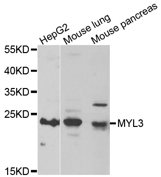 WesternBlot (WB) analysis of MYL3 polyclonal antibody
Bioworld Biotech only provide peptides for our antibodies and do not provide additional peptide customization services.
Price/Size :
USD 368/1mg/vial

Tips:
For phospho antibody, we provide phospho peptide(0.5mg) and non-phospho peptide(0.5mg).Describe :
Blocking peptides are peptides that bind specifically to the target antibody and block antibody binding. These peptide usually contains the epitope recognized by the antibody. Antibodies bound to the blocking peptide no longer bind to the epitope on the target protein. This mechanism is useful when non-specific binding is an issue, for example, in Western blotting (WB) and Immunohistochemistry (IHC). By comparing the staining from the blocked antibody versus the antibody alone, one can see which staining is specific; Specific binding will be absent from the western blot or IHC performed with the neutralized antibody.Formula:
Synthetic peptide was lyophilized with 100% acetonitrile and is supplied as a powder. Reconstitute with 0.1 ml DI water for a final concentration of 10 mg/ml.The purity is >90%,tested by HPLC and MS.
Storage:
The freeze-dried powder is more stable. For short time at 2-8°C. For long term storage store at -20°C.
Note :
This product is for research use only (RUO only). Not for use in diagnostic or therapeutic procedures.