
 DRD1 polyclonal antibody
  Datasheet
 COA
COA MSDS
MSDS SHIP
SHIP
Product Name :
DRD1 polyclonal antibody Background :
The members of the G protein coupled receptor family are distinguished by their slow transmitting response to ligand binding. These transmembrane proteins include the adrenergic, serotonin and dopamine receptors. The effect of the signaling molecule can be excitatory or inhibitory depending on the type of receptor to which it binds. β-adrenergic receptor binds to adrenaline activates adenylyl cyclase, while α2-adrenergic receptor binds to adrenaline inhibits adenylyl cyclase. The dopamine receptors are divided into two classes, D1 and D2, which differ in their functional characteristics in that D1 receptors stimulate adenylyl cyclase while D2 receptors inhibit adenylyl cyclase activity. Five different subtypes of dopamine receptor have been described to date. D1DR and D5DR belong to the D1 subclass, while D2DR, D3DR and D4DR belong to the D2 subclass. Product :
Rabbit IgG, 1mg/ml in PBS with 0.02% sodium azide, 50% glycerol, pH7.2 Storage&Stability :
Store at +4°C after thawing. Aliquot store at -20°C or -80°C. Avoid repeated freeze / thaw cycles. Specificity :
DRD1 polyclonal antibody detects endogenous levels of DRD1 protein. Immunogen :
recombinant protein Conjugate :
Unconjugated Modification :
Unmodification
-
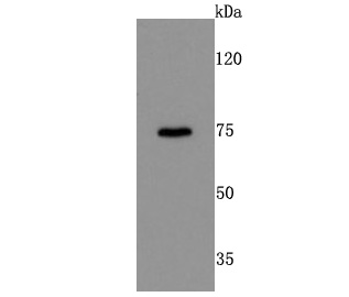 Western blot analysis of DRD1 on mouse kidney cells lysates using anti-DRD1 antibody at 1/500 dilution.
 -
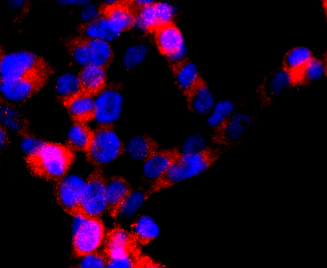 ICC staining DRD1 in 293T cells (red). The nuclear counter stain is DAPI (blue). Cells were fixed in paraformaldehyde, permeabilised with 0.25% Triton X100/PBS.
Bioworld Biotech only provide peptides for our antibodies and do not provide additional peptide customization services.
Price/Size :
USD 368/1mg/vial

Tips:
For phospho antibody, we provide phospho peptide(0.5mg) and non-phospho peptide(0.5mg).Describe :
Blocking peptides are peptides that bind specifically to the target antibody and block antibody binding. These peptide usually contains the epitope recognized by the antibody. Antibodies bound to the blocking peptide no longer bind to the epitope on the target protein. This mechanism is useful when non-specific binding is an issue, for example, in Western blotting (WB) and Immunohistochemistry (IHC). By comparing the staining from the blocked antibody versus the antibody alone, one can see which staining is specific; Specific binding will be absent from the western blot or IHC performed with the neutralized antibody.Formula:
Synthetic peptide was lyophilized with 100% acetonitrile and is supplied as a powder. Reconstitute with 0.1 ml DI water for a final concentration of 10 mg/ml.The purity is >90%,tested by HPLC and MS.
Storage:
The freeze-dried powder is more stable. For short time at 2-8°C. For long term storage store at -20°C.
Note :
This product is for research use only (RUO only). Not for use in diagnostic or therapeutic procedures.