
 EB3 polyclonal antibody
  Datasheet
 COA
COA MSDS
MSDS SHIP
SHIP
Product Name :
EB3 polyclonal antibody Background :
EB1 (MAPRE2, microtubule-associated protein, RP/EB family, member 2, EB2, RP1) may influence tumorigenesis of colorectal cancers and proliferative control of normal cells. EB1 may belong to the intermediate/early gene family, involved in the signal transduction cascade downstream of the TCR. Colorectal cancer is caused by the pathologic transformation of normal colonic epithelium to an adenomatous polyp, which can become an invasive cancer. APC (adenomatous polyposis coli) is a tumor suppressor gene, the mutation of which is one of the earliest events in colorectal carcinogenesis. A majority of the mutations result in the loss of the carboxy terminus of APC. EB1 has been shown to bind to the carboxy terminal region of APC, which implicates EB1 in APC suppression of colonic cancer. EB1 overexpression may play a role in the development of human esophageal squamous cell carcinoma (ESCC) by affecting APC function and activating the beta-catenin/TCF pathway. EB3 is related to EB1 and likewise associates with the microtubule cytoskeleton. EB3 is expressed predominantly in the central nervous system and preferentially associates with APCL. Product :
Rabbit IgG, 1mg/ml in PBS with 0.02% sodium azide, 50% glycerol, pH7.2 Storage&Stability :
Store at +4°C after thawing. Aliquot store at -20°C or -80°C. Avoid repeated freeze / thaw cycles. Specificity :
EB3 polyclonal antibody detects endogenous levels of EB3 protein. Immunogen :
Recombinant protein Conjugate :
Unconjugated Modification :
Unmodification
-
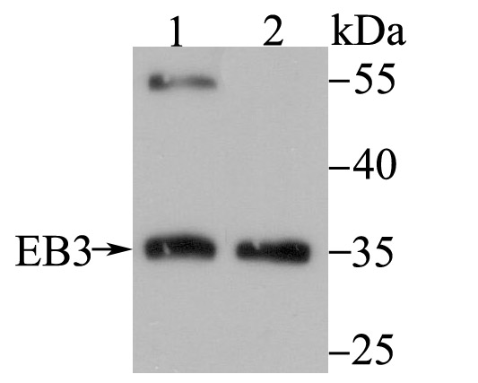 Western blot analysis of EB3 on different lysates using anti-EB3 antibody at 1/500 dilution. Positive control: Lane 1: Mouse brain tissue Lane 2: K562
 -
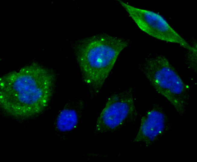 ICC staining EB3 in PC-3M cells (green). The nuclear counter stain is DAPI (blue). Cells were fixed in paraformaldehyde, permeabilised with 0.25% Triton X100/PBS.
Bioworld Biotech only provide peptides for our antibodies and do not provide additional peptide customization services.
Price/Size :
USD 368/1mg/vial

Tips:
For phospho antibody, we provide phospho peptide(0.5mg) and non-phospho peptide(0.5mg).Describe :
Blocking peptides are peptides that bind specifically to the target antibody and block antibody binding. These peptide usually contains the epitope recognized by the antibody. Antibodies bound to the blocking peptide no longer bind to the epitope on the target protein. This mechanism is useful when non-specific binding is an issue, for example, in Western blotting (WB) and Immunohistochemistry (IHC). By comparing the staining from the blocked antibody versus the antibody alone, one can see which staining is specific; Specific binding will be absent from the western blot or IHC performed with the neutralized antibody.Formula:
Synthetic peptide was lyophilized with 100% acetonitrile and is supplied as a powder. Reconstitute with 0.1 ml DI water for a final concentration of 10 mg/ml.The purity is >90%,tested by HPLC and MS.
Storage:
The freeze-dried powder is more stable. For short time at 2-8°C. For long term storage store at -20°C.
Note :
This product is for research use only (RUO only). Not for use in diagnostic or therapeutic procedures.