
 EDG1 polyclonal antibody
  Datasheet
 COA
COA MSDS
MSDS SHIP
SHIP
Product Name :
EDG1 polyclonal antibody Background :
The EDG (endothelial differentiation gene) family of G protein coupled receptors consists of eight family members that bind lysophospholipid (LPL) mediators, including sphingosine-1-phosphate (SPP) and lysophosphatidic acid (LPA). EDG-1, EDG-3, EDG-5 (also designated H218 and AGR16) and EDG-8 bind SPP with high affinity. EDG-6 is a low affinity receptor for SPP. LPA preferentially binds to EDG-2, EDG-4 and EDG-7. The EDG receptors couple to multiple G proteins to signal through Ras, MAP kinase, Rho, Phospholipase C or other tyrosine kinases, which lead to cell survival, growth, migration and differentiation. EDG-1 signals through Gi proteins to activate Akt and is expressed in glioma cells. EDG-2 is expressed in brain, especially in white matter tract regions, while EDG-3 is expressed in cardiovascular tissue and in cerebellum. EDG-4 is highly expressed on leukocytes and brain, and EDG-5 has wide tissue distribution, including cardiovascular tissue and brain. EDG-6, which is expressed in lymphoid and hematopoietic tissues and in lung, signals through G(i/o) proteins, which activate growth related pathways Product :
Rabbit IgG, 1mg/ml in PBS with 0.02% sodium azide, 50% glycerol, pH7.2 Storage&Stability :
Store at +4°C after thawing. Aliquot store at -20°C or -80°C. Avoid repeated freeze / thaw cycles. Specificity :
EDG1 polyclonal antibody detects endogenous levels of EDG1 protein. Immunogen :
recombinant protein Conjugate :
Unconjugated Modification :
Unmodification
-
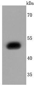 Western blot analysis of EDG1 on SH-SY5Y cells lysates using anti-EDG1 antibody at 1/1,000 dilution.
 -
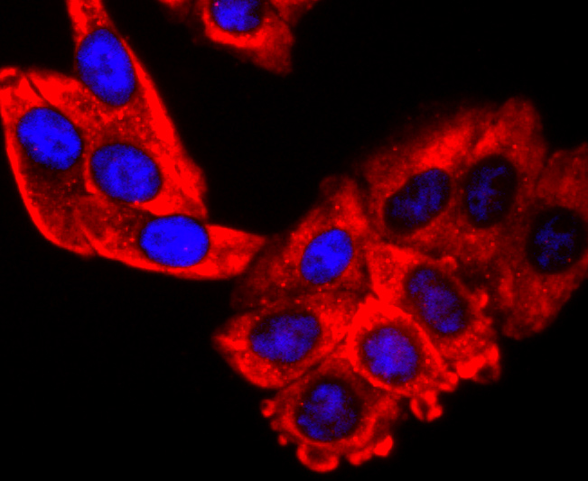 ICC staining EDG1 in HepG2 cells (red). The nuclear counter stain is DAPI (blue). Cells were fixed in paraformaldehyde, permeabilised with 0.25% Triton X100/PBS.
Bioworld Biotech only provide peptides for our antibodies and do not provide additional peptide customization services.
Price/Size :
USD 368/1mg/vial

Tips:
For phospho antibody, we provide phospho peptide(0.5mg) and non-phospho peptide(0.5mg).Describe :
Blocking peptides are peptides that bind specifically to the target antibody and block antibody binding. These peptide usually contains the epitope recognized by the antibody. Antibodies bound to the blocking peptide no longer bind to the epitope on the target protein. This mechanism is useful when non-specific binding is an issue, for example, in Western blotting (WB) and Immunohistochemistry (IHC). By comparing the staining from the blocked antibody versus the antibody alone, one can see which staining is specific; Specific binding will be absent from the western blot or IHC performed with the neutralized antibody.Formula:
Synthetic peptide was lyophilized with 100% acetonitrile and is supplied as a powder. Reconstitute with 0.1 ml DI water for a final concentration of 10 mg/ml.The purity is >90%,tested by HPLC and MS.
Storage:
The freeze-dried powder is more stable. For short time at 2-8°C. For long term storage store at -20°C.
Note :
This product is for research use only (RUO only). Not for use in diagnostic or therapeutic procedures.