
 Hip1 polyclonal antibody
  Datasheet
 COA
COA MSDS
MSDS SHIP
SHIP
Product Name :
Hip1 polyclonal antibody Background :
Huntington disease is associated with the expansion of a polyglutamine tract, greater than 35 repeats, in the HD gene product huntingtin. HIP1 (huntingtin-interacting protein 1), a membrane-associated protein, binds specifically to the N-terminus of human huntingtin. HIP1 is ubiquitously expressed in different brain regions at low levels, and exhibits nearly identical subcellular fractionation as huntingtin. The huntingtin-HIP1 interaction is restricted to the brain and is inversely correlated to the polyglutamine length in the huntingtin, suggesting that loss of normal huntingtin-HIP1 interaction may compromise the membrane-cytoskeletal integrity in the brain. HIP1 contains an endocytic multidomain protein with a C-terminal Actin-binding domain, a central coiled-coil forming region and an N-terminal ENTH domain. HIP1 may be involved in vesicle trafficking; the structural integrity of HIP1 is crucial for maintenance of normal vesicle size in vivo. HIP12 is a non-proapoptotic member of the HIP gene family that is expressed in the brain and shares a similar subcellular distribution pattern with HIP1. However, HIP12 differs from HIP1 in its pattern of expression at both the mRNA and protein level. HIP12 does not directly interact with huntingtin but can interact with HIP1. Product :
Rabbit IgG, 1mg/ml in PBS with 0.02% sodium azide, 50% glycerol, pH7.2 Storage&Stability :
Store at +4°C after thawing. Aliquot store at -20°C. Avoid repeated freeze / thaw cycles. Specificity :
Hip1 polyclonal antibody detects endogenous levels of Hip1 protein. Immunogen :
Synthetic peptide within C terminal human Hip1. Conjugate :
Unconjugated Modification :
Unmodification
-
 Western blot analysis of Hip1 on different tissue lysates using anti-Hip1 antibody at 1/500 dilution. Positive control: Lane1: Mouse testis Lane2: Mouse spinal cord Lane3: SH-SY5Y
 -
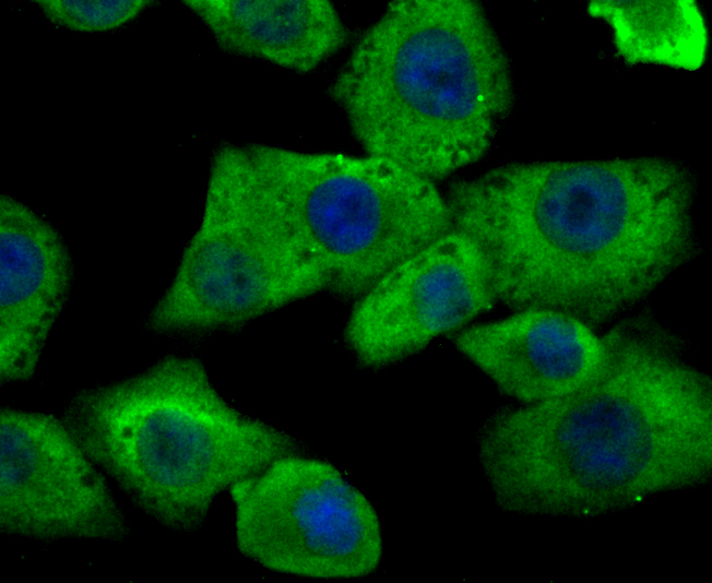 ICC staining Hip1 in A549 cells (green). The nuclear counter stain is DAPI (blue). Cells were fixed in paraformaldehyde, permeabilised with 0.25% Triton X100/PBS.
Bioworld Biotech only provide peptides for our antibodies and do not provide additional peptide customization services.
Price/Size :
USD 368/1mg/vial

Tips:
For phospho antibody, we provide phospho peptide(0.5mg) and non-phospho peptide(0.5mg).Describe :
Blocking peptides are peptides that bind specifically to the target antibody and block antibody binding. These peptide usually contains the epitope recognized by the antibody. Antibodies bound to the blocking peptide no longer bind to the epitope on the target protein. This mechanism is useful when non-specific binding is an issue, for example, in Western blotting (WB) and Immunohistochemistry (IHC). By comparing the staining from the blocked antibody versus the antibody alone, one can see which staining is specific; Specific binding will be absent from the western blot or IHC performed with the neutralized antibody.Formula:
Synthetic peptide was lyophilized with 100% acetonitrile and is supplied as a powder. Reconstitute with 0.1 ml DI water for a final concentration of 10 mg/ml.The purity is >90%,tested by HPLC and MS.
Storage:
The freeze-dried powder is more stable. For short time at 2-8°C. For long term storage store at -20°C.
Note :
This product is for research use only (RUO only). Not for use in diagnostic or therapeutic procedures.