
 MLH1 polyclonal antibody
  Datasheet
 COA
COA MSDS
MSDS SHIP
SHIP
Product Name :
MLH1 polyclonal antibody Background :
DNA-mismatch repair (MMR) is an essential process in maintaining genetic stability. Lack of a functional DNA-mismatch repair pathway is a common characteristic of several different types of human cancers, either due to an MMR gene mutation or promoter methylation gene silencing. MLH1 is an integral part of the protein complex responsible for mismatch repair and is expressed in lymphocytes, heart, colon, breast, lung, spleen, testis, prostate, thyroid and gall bladder tissues, and is methylated in several ovarian tumors. Loss of MLH1 protein expression is associated with a mutated phenotype, microsatellite instability and a predisposition to cancer. In hereditary nonpolyposis colorectal cancer (HNPCC), an autosomal dominant inherited cancer syndrome that signifies a high risk of colorectal and various other types of cancer, the MLH1 gene exhibits a pathogenic mutation. Certain cancer cell lines, including leukemia CCRF-CEM, colon HCT 116 and KM12, and ovarian cancers SK-OV-3 and IGROV-1, show complete deficiency of MLH1, while MLH1 is expressed in 60% of melanomas, 70% of noninvasive squamous cell carcinomas and 30% of invasive squamous cell carcinomas. Product :
Rabbit IgG, 1mg/ml in PBS with 0.02% sodium azide, 50% glycerol, pH7.2 Storage&Stability :
Store at +4°C after thawing. Aliquot store at -20°C or -80°C. Avoid repeated freeze / thaw cycles. Specificity :
MLH1 polyclonal antibody detects endogenous levels of MLH1 protein. Immunogen :
recombinant protein Conjugate :
Unconjugated Modification :
Unmodification
-
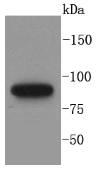 Western blot analysis of MLH1 on A431 cells lysates using anti-MLH1 antibody at 1/1,000 dilution.
 -
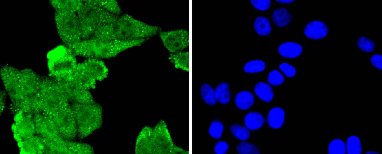 ICC staining MLH1 in HepG2 cells (green). The nuclear counter stain is DAPI (blue). Cells were fixed in paraformaldehyde, permeabilised with 0.25% Triton X100/PBS.
Bioworld Biotech only provide peptides for our antibodies and do not provide additional peptide customization services.
Price/Size :
USD 368/1mg/vial

Tips:
For phospho antibody, we provide phospho peptide(0.5mg) and non-phospho peptide(0.5mg).Describe :
Blocking peptides are peptides that bind specifically to the target antibody and block antibody binding. These peptide usually contains the epitope recognized by the antibody. Antibodies bound to the blocking peptide no longer bind to the epitope on the target protein. This mechanism is useful when non-specific binding is an issue, for example, in Western blotting (WB) and Immunohistochemistry (IHC). By comparing the staining from the blocked antibody versus the antibody alone, one can see which staining is specific; Specific binding will be absent from the western blot or IHC performed with the neutralized antibody.Formula:
Synthetic peptide was lyophilized with 100% acetonitrile and is supplied as a powder. Reconstitute with 0.1 ml DI water for a final concentration of 10 mg/ml.The purity is >90%,tested by HPLC and MS.
Storage:
The freeze-dried powder is more stable. For short time at 2-8°C. For long term storage store at -20°C.
Note :
This product is for research use only (RUO only). Not for use in diagnostic or therapeutic procedures.