
 mSin3A polyclonal antibody
  Datasheet
 COA
COA MSDS
MSDS SHIP
SHIP
Product Name :
mSin3A polyclonal antibody Background :
It is now well established that Myc regulation of cell proliferation and differentiation involves a family of related transcription factors. One such factor, Max, is an obligate heterodimeric partner for Myc and can also form heterodimers with at least four related proteins designated Mad 1, Mxi1 (alternatively designated Mad 2), Mad 3 and Mad 4. Like Mad 1 and Mxi1, association of Mad 3 and Mad 4 with Max results in transcriptional repression. Both Myc and the Mad proteins have short half-lives and their synthesis is tightly regulated, while Max expression is constitutive and relatively stable. Two related mammalian cDNAs have been identified and shown to encode Mad-binding proteins. Both possess sequence homology with the yeast transcription repressor Sin3 including four conserved paired amphipathic helix (PAH) domains. mSin3A and mSin3B specifically interact with the Mad proteins via their second paired amphipathic helix domain (PAH2). It has been suggested that Mad-Max heterodimers repress transcription by tethering mSin3 to DNA as corepressors. Product :
Rabbit IgG, 1mg/ml in PBS with 0.02% sodium azide, 50% glycerol, pH7.2 Storage&Stability :
Store at +4°C after thawing. Aliquot store at -20°C or -80°C. Avoid repeated freeze / thaw cycles. Specificity :
mSin3A polyclonal antibody detects endogenous levels of mSin3A protein. Immunogen :
recombinant protein Conjugate :
Unconjugated Modification :
Unmodification
-
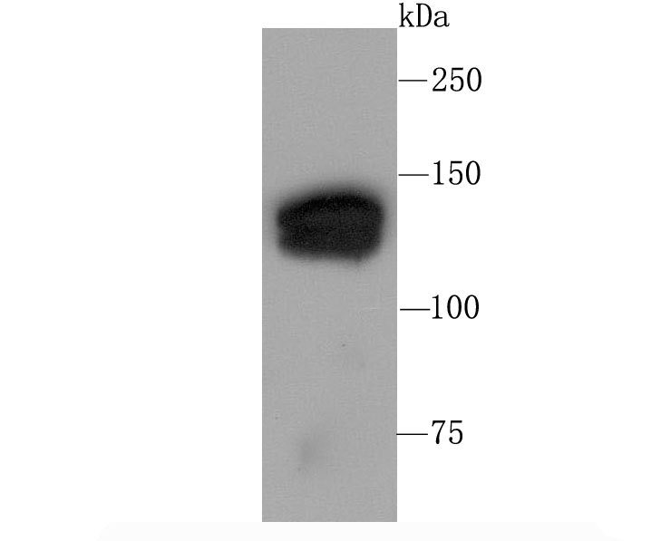 Western blot analysis of mSin3A on 293T cell using anti-mSin3A antibody at 1/1,000 dilution.
 -
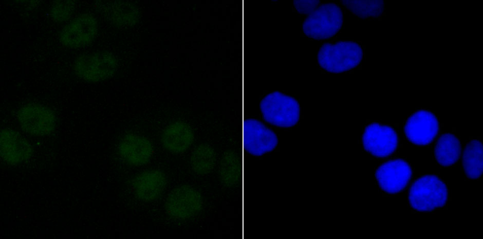 ICC staining mSin3A in Hela cells (green). The nuclear counter stain is DAPI (blue). Cells were fixed in paraformaldehyde, permeabilised with 0.25% Triton X100/PBS.
Bioworld Biotech only provide peptides for our antibodies and do not provide additional peptide customization services.
Price/Size :
USD 368/1mg/vial

Tips:
For phospho antibody, we provide phospho peptide(0.5mg) and non-phospho peptide(0.5mg).Describe :
Blocking peptides are peptides that bind specifically to the target antibody and block antibody binding. These peptide usually contains the epitope recognized by the antibody. Antibodies bound to the blocking peptide no longer bind to the epitope on the target protein. This mechanism is useful when non-specific binding is an issue, for example, in Western blotting (WB) and Immunohistochemistry (IHC). By comparing the staining from the blocked antibody versus the antibody alone, one can see which staining is specific; Specific binding will be absent from the western blot or IHC performed with the neutralized antibody.Formula:
Synthetic peptide was lyophilized with 100% acetonitrile and is supplied as a powder. Reconstitute with 0.1 ml DI water for a final concentration of 10 mg/ml.The purity is >90%,tested by HPLC and MS.
Storage:
The freeze-dried powder is more stable. For short time at 2-8°C. For long term storage store at -20°C.
Note :
This product is for research use only (RUO only). Not for use in diagnostic or therapeutic procedures.