
 Ndufs4 polyclonal antibody
  Datasheet
 COA
COA MSDS
MSDS SHIP
SHIP
Product Name :
Ndufs4 polyclonal antibody Background :
NADH dehydrogenase [ubiquinone] iron-sulfur protein 4, mitochondrial (NDUFS4) also known as NADH-ubiquinone oxidoreductase 18 kDa subunit is an enzyme that in humans is encoded by the NDUFS4 gene. This gene encodes an nuclear-encoded accessory subunit of the mitochondrial membrane respiratory chain NADH dehydrogenase (complex I, or NADH:ubiquinone oxidoreductase), plays a vital role in cellular ATP production, the primary source of energy for many crucial processes in living cells. It removes electrons from NADH and passes them by a series of different protein-coupled redox centers to the electron acceptor ubiquinone. In well-coupled mitochondria, the electron flux leads to ATP generation via the building of a proton gradient across the inner membrane. Complex I is composed of at least 41 subunits, of which 7 are encoded by the mitochondrial genome (ND1-6, ND4L) and the remainder by nuclear genes. Mutations in this gene can cause mitochondrial complex I deficiencies such as Leigh syndrome. Product :
Rabbit IgG, 1mg/ml in PBS with 0.02% sodium azide, 50% glycerol, pH7.2 Storage&Stability :
Store at +4℃ after thawing. Aliquot store at -20℃. Avoid repeated freeze / thaw cycles. Specificity :
Ndufs4 polyclonal antibody detects endogenous levels of Ndufs4 protein. Immunogen :
Full length recombinant protein. Conjugate :
Unconjugated Modification :
Unmodification
-
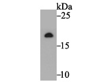 Western blot analysis of Ndufs4 on rat heart tissue lysate. Proteins were transferred to a PVDF membrane and blocked with 5% BSA in PBS for 1 hour at room temperature. The primary antibody was used at a 1:500 dilution in 5% BSA at room temperature for 2 hours. Goat Anti-Rabbit IgG - HRP Secondary Antibody (HA1001) at 1:5,000 dilution was used for 1 hour at room temperature.
 -
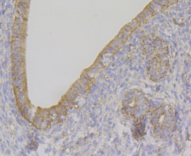 Immunohistochemical analysis of paraffin-embedded rat uterus tissue using anti-Ndufs4 antibody. The section was pre-treated using heat mediated antigen retrieval with Tris-EDTA buffer (pH 8.0-8.4) for 20 minutes. The tissues were blocked in 5% BSA for 30 minutes at room temperature, washed with ddH2O and PBS, and then probed with ET7109-37 at 1/100 dilution, for 30 minutes at room temperature and detected using an HRP conjugated compact polymer system. DAB was used as the chrogen. Counter stained with hematoxylin and mounted with DPX.
Bioworld Biotech only provide peptides for our antibodies and do not provide additional peptide customization services.
Price/Size :
USD 368/1mg/vial

Tips:
For phospho antibody, we provide phospho peptide(0.5mg) and non-phospho peptide(0.5mg).Describe :
Blocking peptides are peptides that bind specifically to the target antibody and block antibody binding. These peptide usually contains the epitope recognized by the antibody. Antibodies bound to the blocking peptide no longer bind to the epitope on the target protein. This mechanism is useful when non-specific binding is an issue, for example, in Western blotting (WB) and Immunohistochemistry (IHC). By comparing the staining from the blocked antibody versus the antibody alone, one can see which staining is specific; Specific binding will be absent from the western blot or IHC performed with the neutralized antibody.Formula:
Synthetic peptide was lyophilized with 100% acetonitrile and is supplied as a powder. Reconstitute with 0.1 ml DI water for a final concentration of 10 mg/ml.The purity is >90%,tested by HPLC and MS.
Storage:
The freeze-dried powder is more stable. For short time at 2-8°C. For long term storage store at -20°C.
Note :
This product is for research use only (RUO only). Not for use in diagnostic or therapeutic procedures.