
 Perforin polyclonal antibody
  Datasheet
 COA
COA MSDS
MSDS SHIP
SHIP
Product Name :
Perforin polyclonal antibody Background :
The protein encoded by this gene has structural and functional similarities to complement component 9 (C9). Like C9, this protein creates transmembrane tubules and is capable of lysing non-specifically a variety of target cells. Plays a key role in secretory granule-dependent cell death, and in defense against virus-infected or neoplastic cells. This protein is one of the main cytolytic proteins of cytolytic granules, and it is known to be a key effector molecule for T-cell- and natural killer-cell-mediated cytolysis. Defects in this gene cause familial hemophagocytic lymphohistiocytosis type 2 (HPLH2), a rare and lethal autosomal recessive disorder of early childhood. Alternative splicing results in multiple transcript variants encoding the same protein. Plays an important role in killing other cells that are recognized as non-self by the immune system, e.g. in transplant rejection or some forms of autoimmune disease. Can insert into the membrane of target cells in its calcium-bound form, oligomerize and form large pores. Product :
Rabbit IgG, 1mg/ml in PBS with 0.02% sodium azide, 50% glycerol, pH7.2 Storage&Stability :
Store at +4℃ after thawing. Aliquot store at -20℃. Avoid repeated freeze / thaw cycles. Specificity :
Perforin polyclonal antibody detects endogenous levels of Perforin protein. Immunogen :
Recombinant protein within Human Perforin aa 300-500. Conjugate :
Unconjugated Modification :
Unmodification
-
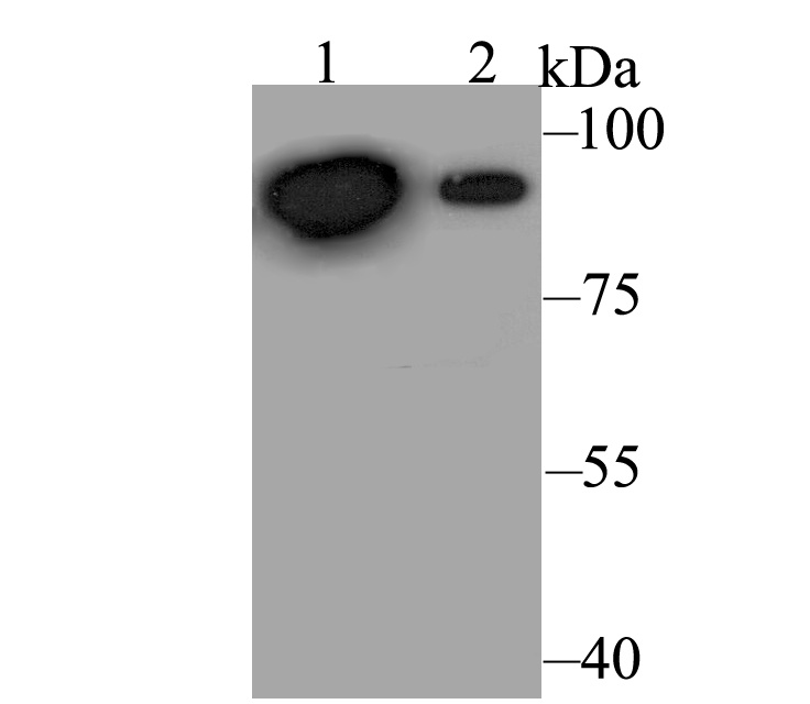 Western blot analysis of Perforin on different lysates. Proteins were transferred to a PVDF membrane and blocked with 5% BSA in PBS for 1 hour at room temperature. The primary antibody was used at a 1:500 dilution in 5% BSA at room temperature for 2 hours. Goat Anti-Rabbit IgG - HRP Secondary Antibody (HA1001) at 1:5,000 dilution was used for 1 hour at room temperature.Positive control: Lane 1: HepG2 cell lysateLane 2: Daudi cell lysate
 -
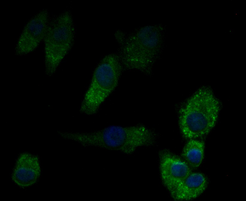 ICC staining Perforin in A549 cells (green). Formalin fixed cells were permeabilized with 0.1% Triton X-100 in TBS for 10 minutes at room temperature and blocked with 1% Blocker BSA for 15 minutes at room temperature. Cells were probed with Perforin polyclonal antibody at a dilution of 1:200 for 1 hour at room temperature, washed with PBS. Alexa Fluorc™ 488 Goat anti-Rabbit IgG was used as the secondary antibody at 1/100 dilution. The nuclear counter stain is DAPI (blue).
Bioworld Biotech only provide peptides for our antibodies and do not provide additional peptide customization services.
Price/Size :
USD 368/1mg/vial

Tips:
For phospho antibody, we provide phospho peptide(0.5mg) and non-phospho peptide(0.5mg).Describe :
Blocking peptides are peptides that bind specifically to the target antibody and block antibody binding. These peptide usually contains the epitope recognized by the antibody. Antibodies bound to the blocking peptide no longer bind to the epitope on the target protein. This mechanism is useful when non-specific binding is an issue, for example, in Western blotting (WB) and Immunohistochemistry (IHC). By comparing the staining from the blocked antibody versus the antibody alone, one can see which staining is specific; Specific binding will be absent from the western blot or IHC performed with the neutralized antibody.Formula:
Synthetic peptide was lyophilized with 100% acetonitrile and is supplied as a powder. Reconstitute with 0.1 ml DI water for a final concentration of 10 mg/ml.The purity is >90%,tested by HPLC and MS.
Storage:
The freeze-dried powder is more stable. For short time at 2-8°C. For long term storage store at -20°C.
Note :
This product is for research use only (RUO only). Not for use in diagnostic or therapeutic procedures.