
 RNF40 polyclonal antibody
  Datasheet
 COA
COA MSDS
MSDS SHIP
SHIP
Product Name :
RNF40 polyclonal antibody Background :
Component of the RNF20/40 E3 ubiquitin-protein ligase complex that mediates monoubiquitination of 'Lys-120' of histone H2B (H2BK120ub1). H2BK120ub1 gives a specific tag for epigenetic transcriptional activation and is also prerequisite for histone H3 'Lys-4' and 'Lys-79' methylation (H3K4me and H3K79me, respectively). It thereby plays a central role in histone code and gene regulation. The RNF20/40 complex forms a H2B ubiquitin ligase complex in cooperation with the E2 enzyme UBE2A or UBE2B; reports about the cooperation with UBE2E1/UBCH are contradictory. Required for transcriptional activation of Hox genes. This protein was reported to interact with the tumor suppressor protein RB1. Studies of the rat counterpart suggested that this protein may function as an E3 ubiquitin-protein ligase, and facilitate the ubiquitination and degradation of syntaxin 1, which is an essential component of the neurotransmitter release machinery. Multiple alternatively spliced transcript variants encoding different isoforms have been found for this gene. Product :
Rabbit IgG, 1mg/ml in PBS with 0.02% sodium azide, 50% glycerol, pH7.2 Storage&Stability :
Store at +4℃ after thawing. Aliquot store at -20℃. Avoid repeated freeze / thaw cycles. Specificity :
RNF40 polyclonal antibody detects endogenous levels of RNF40 protein. Immunogen :
Recombinant protein within human RNF40 aa 200-400. Conjugate :
Unconjugated Modification :
Unmodification
-
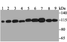 Western blot analysis of RNF40 on different lysates. Proteins were transferred to a PVDF membrane and blocked with 5% BSA in PBS for 1 hour at room temperature. The primary antibody was used at a 1:500 dilution in 5% BSA at room temperature for 2 hours. Goat Anti-Rabbit IgG - HRP Secondary Antibody (HA1001) at 1:5,000 dilution was used for 1 hour at room temperature.Positive control: Lane 1: Mouse testis tissue lysateLane 2: Rat brain tissue lysateLane 3: A431 cell lysateLane 4: SH-SY-5Y cell ly
 -
 Immunohistochemical analysis of paraffin-embedded human tonsil tissue using anti-RNF40 antibody. The section was pre-treated using heat mediated antigen retrieval with sodium citrate buffer (pH 6.0) for 20 mins. The tissues were blocked in 5% BSA for 30 minutes at room temperature, washed with ddH2O and PBS, and then probed with ET7109-46 at 1/50 dilution, for 30 minutes at room temperature and detected using an HRP conjugated compact polymer system. DAB was used as the chrogen. Counter stained with hematoxylin and mounted with DPX.
Bioworld Biotech only provide peptides for our antibodies and do not provide additional peptide customization services.
Price/Size :
USD 368/1mg/vial

Tips:
For phospho antibody, we provide phospho peptide(0.5mg) and non-phospho peptide(0.5mg).Describe :
Blocking peptides are peptides that bind specifically to the target antibody and block antibody binding. These peptide usually contains the epitope recognized by the antibody. Antibodies bound to the blocking peptide no longer bind to the epitope on the target protein. This mechanism is useful when non-specific binding is an issue, for example, in Western blotting (WB) and Immunohistochemistry (IHC). By comparing the staining from the blocked antibody versus the antibody alone, one can see which staining is specific; Specific binding will be absent from the western blot or IHC performed with the neutralized antibody.Formula:
Synthetic peptide was lyophilized with 100% acetonitrile and is supplied as a powder. Reconstitute with 0.1 ml DI water for a final concentration of 10 mg/ml.The purity is >90%,tested by HPLC and MS.
Storage:
The freeze-dried powder is more stable. For short time at 2-8°C. For long term storage store at -20°C.
Note :
This product is for research use only (RUO only). Not for use in diagnostic or therapeutic procedures.