
 RUNX1/2/3 polyclonal antibody
  Datasheet
 COA
COA MSDS
MSDS SHIP
SHIP
Product Name :
RUNX1/2/3 polyclonal antibody Background :
The mammalian Runt-related transcription factor (RUNX) family comprises three members, RUNX1 (also designated AML-1, PEBP2αB, CBFA2), RUNX2 (also designated AML-3, PEBP2αA, CBFA1, Osf2) and RUNX3 (also designated AML-2, PEBPαC, CBFA3). RUNX family members are DNA-binding proteins that regulate the expression of genes involved in cellular differentiation and cell cycle progression. RUNX1 is involved in hematopoiesis and is frequently targeted in human leukemia by chromosomal translocations that fuse the DNA-binding domain of RUNX1 to other transcription factors and corepressor molecules. In addition to its role in leukemogenesis, RUNX1 is also involved in sensory neuron diversification. RUNX1 promotes axonal growth, is selectively expressed in neural crest-derived TrkA+ sensory neurons and mediates TrkA transactivation in migratory neural crest cells. RUNX2 is essential for skeletal mineralization in that it stimulates osteoblast differentiation of mesenchymal stem cells, promotes chondrocyte hypertrophy and contributes to endothelial cell migration and vascular invasion of developing bones. Regulating RUNX2 expression may be a useful therapeutic tool for promoting bone formation. Mutations in the C-terminus of RUNX2 are associated with cleidocranial dysplasia syndrome, an autosomal-dominant skeletal dysplasia syndrome that is characterized by widely patent calvarial sutures, clavicular hypoplasia, supernumerary teeth, and short stature. RUNX3 is expressed in cells of hematopoietic origin, including myeloid and B-cell lines and spleen. By playing a role in controlling the growth and differentiation of gastric epithelial cells, RUNX3 is a strong candidate as a gastric cancer tumor suppressor. Specifically, hypermethylation inactivates the gene encoding RUNX3. The detection of hypermethylation at multiple regions within the RUNX3 CpG island may aid in the diagnosis and risk assessment of gastric cancer. Product :
Rabbit IgG, 1mg/ml in PBS with 0.02% sodium azide, 50% glycerol, pH7.2 Storage&Stability :
Store at +4°C after thawing. Aliquot store at -20°C or -80°C. Avoid repeated freeze / thaw cycles. Specificity :
RUNX1/2/3 polyclonal antibody detects endogenous levels of RUNX1/2/3 protein. Immunogen :
recombinant protein Conjugate :
Unconjugated Modification :
Unmodification
-
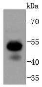 Western blot analysis of RUNX1+RUNX2+RUNX3 on Jurkat cells lysates using anti-RUNX1+RUNX2+RUNX3 antibody at 1/1,000 dilution.
 -
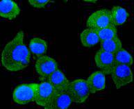 ICC staining RUNX1+RUNX2+RUNX3 in N2A cells (green). The nuclear counter stain is DAPI (blue). Cells were fixed in paraformaldehyde, permeabilised with 0.25% Triton X100/PBS.
Bioworld Biotech only provide peptides for our antibodies and do not provide additional peptide customization services.
Price/Size :
USD 368/1mg/vial

Tips:
For phospho antibody, we provide phospho peptide(0.5mg) and non-phospho peptide(0.5mg).Describe :
Blocking peptides are peptides that bind specifically to the target antibody and block antibody binding. These peptide usually contains the epitope recognized by the antibody. Antibodies bound to the blocking peptide no longer bind to the epitope on the target protein. This mechanism is useful when non-specific binding is an issue, for example, in Western blotting (WB) and Immunohistochemistry (IHC). By comparing the staining from the blocked antibody versus the antibody alone, one can see which staining is specific; Specific binding will be absent from the western blot or IHC performed with the neutralized antibody.Formula:
Synthetic peptide was lyophilized with 100% acetonitrile and is supplied as a powder. Reconstitute with 0.1 ml DI water for a final concentration of 10 mg/ml.The purity is >90%,tested by HPLC and MS.
Storage:
The freeze-dried powder is more stable. For short time at 2-8°C. For long term storage store at -20°C.
Note :
This product is for research use only (RUO only). Not for use in diagnostic or therapeutic procedures.