
 S100 polyclonal antibody
  Datasheet
 COA
COA MSDS
MSDS SHIP
SHIP
Product Name :
S100 polyclonal antibody Background :
The family of EF-hand type Ca2+-binding proteins includes calbindin (previously designated vitamin D-dependent Ca2+-binding protein), S-100 α and β, calgranulins A (also designated MRP8), B (also designated MRP14) and C (S-100 like proteins), and the parvalbumin family members, including parvalbumin α and parvalbumin β (also designated oncomodulin). The S-100 protein is involved in the regulation of cellular processes such as cell cycle progression and differentiation. Research also indicates that the S-100 protein may function in the activation of Ca2+ induced Ca2+ release, inhibition of microtubule assembly and inhibition of protein kinase C mediated phosphorylation. Two S-100 subunits, sharing 60% sequence identity, have been described as S-100 α chain and S-100 β chain. Three S-100 dimeric forms have been characterized, differing in their subunit composition of either two α chains, two β chains or one α and one β chain. S-100 localizes to the cytoplasm and nuclei of astrocytes, Schwann's cells, ependymomas and astrogliomas. S-100 is also detected in almost all benign naevi, malignant melanocytic tumours and in Langerhans cells in the skin. Calbindin, S-100 proteins and parvalbumin proteins are each expressed in neural tissues. In addition, S-100 α and β are present in a variety of other tissues, and calbindin is present in intestine and kidney. Product :
Rabbit IgG, 1mg/ml in PBS with 0.02% sodium azide, 50% glycerol, pH7.2 Storage&Stability :
Store at +4°C after thawing. Aliquot store at -20°C or -80°C. Avoid repeated freeze / thaw cycles. Specificity :
S100 polyclonal antibody detects endogenous levels of S100 protein. Immunogen :
recombinant protein Conjugate :
Unconjugated Modification :
Unmodification
-
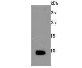 Western blot analysis of S100 on mouse heart cells lysates using anti-S100 antibody at 1/500 dilution.
 -
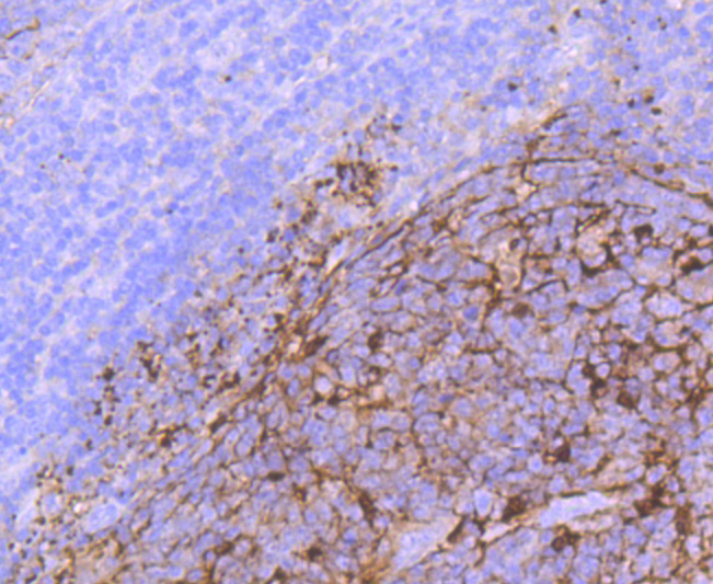 Immunohistochemical analysis of paraffin-embedded human tonsil tissue using anti- S100 antibody. Counter stained with hematoxylin.
Bioworld Biotech only provide peptides for our antibodies and do not provide additional peptide customization services.
Price/Size :
USD 368/1mg/vial

Tips:
For phospho antibody, we provide phospho peptide(0.5mg) and non-phospho peptide(0.5mg).Describe :
Blocking peptides are peptides that bind specifically to the target antibody and block antibody binding. These peptide usually contains the epitope recognized by the antibody. Antibodies bound to the blocking peptide no longer bind to the epitope on the target protein. This mechanism is useful when non-specific binding is an issue, for example, in Western blotting (WB) and Immunohistochemistry (IHC). By comparing the staining from the blocked antibody versus the antibody alone, one can see which staining is specific; Specific binding will be absent from the western blot or IHC performed with the neutralized antibody.Formula:
Synthetic peptide was lyophilized with 100% acetonitrile and is supplied as a powder. Reconstitute with 0.1 ml DI water for a final concentration of 10 mg/ml.The purity is >90%,tested by HPLC and MS.
Storage:
The freeze-dried powder is more stable. For short time at 2-8°C. For long term storage store at -20°C.
Note :
This product is for research use only (RUO only). Not for use in diagnostic or therapeutic procedures.