
 SNX1 polyclonal antibody
  Datasheet
 COA
COA MSDS
MSDS SHIP
SHIP
Product Name :
SNX1 polyclonal antibody Background :
Sorting nexin 1 (SNX1) is a member of a large family of hydrophilic proteins that interact with a variety of receptor types and are involved in intracellular trafficking. SNX1 and the related splice variant, SNX1A, bind the epidermal growth factor (EGF) receptor, facilitate its transport to lysosome, and thereby contribute to the degradation of the receptor. SNX2 and SNX4 share a high degree of amino acid similarity with SNX1, as they all contain a characteristic phox homology (PX) domain. These proteins are all partially associated with cellular membranes, and they, likewise, associate with EGF, PDGF and insulin receptor tyrosine kinases. These nexins are widely expressed and yet have various tissue distribution patterns. Additionally, the sorting nexins can associate with each other and with a variety of other cellular proteins, suggesting that they exist as part of multisubunit complexes. The related protein, SNX3, comprises a distinct subgroup of nexins that share less sequence similarity outside of the PX domain and have dramatically different binding affinities for the tyrosine kinase receptors. Product :
Rabbit IgG, 1mg/ml in PBS with 0.02% sodium azide, 50% glycerol, pH7.2 Storage&Stability :
Store at +4°C after thawing. Aliquot store at -20°C. Avoid repeated freeze / thaw cycles. Specificity :
SNX1 polyclonal antibody detects endogenous levels of SNX1 protein. Immunogen :
Recombinant protein within human SNX1 aa 50-250. Conjugate :
Unconjugated Modification :
Unmodification
-
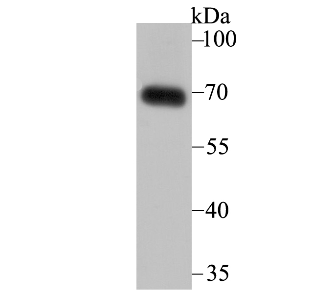 Western blot analysis of SNX1 on human skin tissue lysate using anti-SNX1 antibody at 1/2,000 dilution.
 -
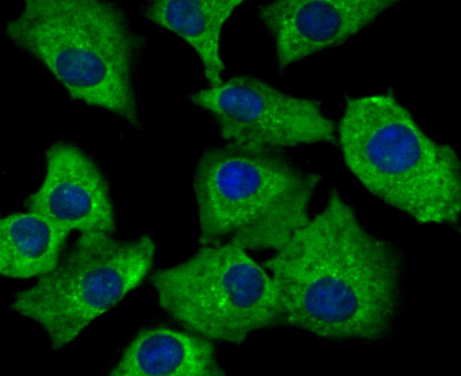 ICC staining SNX1 in A549 cells (green). The nuclear counter stain is DAPI (blue). Cells were fixed in paraformaldehyde, permeabilised with 0.25% Triton X100/PBS.
Bioworld Biotech only provide peptides for our antibodies and do not provide additional peptide customization services.
Price/Size :
USD 368/1mg/vial

Tips:
For phospho antibody, we provide phospho peptide(0.5mg) and non-phospho peptide(0.5mg).Describe :
Blocking peptides are peptides that bind specifically to the target antibody and block antibody binding. These peptide usually contains the epitope recognized by the antibody. Antibodies bound to the blocking peptide no longer bind to the epitope on the target protein. This mechanism is useful when non-specific binding is an issue, for example, in Western blotting (WB) and Immunohistochemistry (IHC). By comparing the staining from the blocked antibody versus the antibody alone, one can see which staining is specific; Specific binding will be absent from the western blot or IHC performed with the neutralized antibody.Formula:
Synthetic peptide was lyophilized with 100% acetonitrile and is supplied as a powder. Reconstitute with 0.1 ml DI water for a final concentration of 10 mg/ml.The purity is >90%,tested by HPLC and MS.
Storage:
The freeze-dried powder is more stable. For short time at 2-8°C. For long term storage store at -20°C.
Note :
This product is for research use only (RUO only). Not for use in diagnostic or therapeutic procedures.