
 TNFR2 polyclonal antibody
  Datasheet
 COA
COA MSDS
MSDS SHIP
SHIP
Product Name :
TNFR2 polyclonal antibody Background :
Tumor necrosis factor (TNF) is a pleiotropic cytokine whose function is mediated through two distinct cell surface receptors. These receptors, designated TNF-R1 and TNF-R2, are expressed on most cell types. The majority of TNF functions are primarily mediated through TNF-R1, while signaling through TNF-R2 occurs less extensively and is confined to cells of the immune system. Both of these proteins belong to the growing TNF and nerve growth factor (NGF) receptor superfamily, which includes FAS, CD30, CD27 and CD40. The members of this superfamily are type I membrane proteins that share sequence homology confined to the extracellular region. TNF-R1 shares a motif termed the "death domain" with FAS and three structurally unrelated signaling proteins, TRADD, FADD and RIP (1,3-8). This death domain is required for transduction of the apoptotic signal. Product :
Rabbit IgG, 1mg/ml in PBS with 0.02% sodium azide, 50% glycerol, pH7.2 Storage&Stability :
Store at +4°C after thawing. Aliquot store at -20°C or -80°C. Avoid repeated freeze / thaw cycles. Specificity :
TNFR2 polyclonal antibody detects endogenous levels of TNFR2 protein. Immunogen :
recombinant protein Conjugate :
Unconjugated Modification :
Unmodification
-
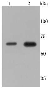 Western blot analysis of TNF Receptor II on different cells lysates using anti-TNF Receptor II antibody at 1/500 dilution. Positive control: Line 1: MCF-7 Line 2: Jurkat
 -
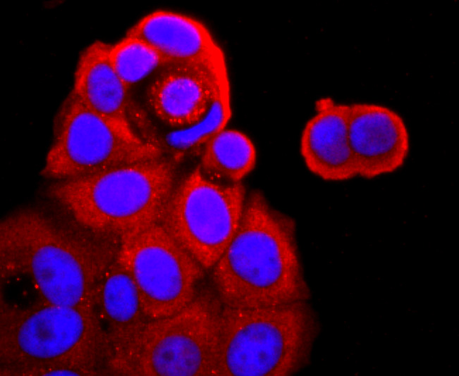 ICC staining TNF Receptor II in MCF-7 cells (red). The nuclear counter stain is DAPI (blue). Cells were fixed in paraformaldehyde, permeabilised with 0.25% Triton X100/PBS.
Bioworld Biotech only provide peptides for our antibodies and do not provide additional peptide customization services.
Price/Size :
USD 368/1mg/vial

Tips:
For phospho antibody, we provide phospho peptide(0.5mg) and non-phospho peptide(0.5mg).Describe :
Blocking peptides are peptides that bind specifically to the target antibody and block antibody binding. These peptide usually contains the epitope recognized by the antibody. Antibodies bound to the blocking peptide no longer bind to the epitope on the target protein. This mechanism is useful when non-specific binding is an issue, for example, in Western blotting (WB) and Immunohistochemistry (IHC). By comparing the staining from the blocked antibody versus the antibody alone, one can see which staining is specific; Specific binding will be absent from the western blot or IHC performed with the neutralized antibody.Formula:
Synthetic peptide was lyophilized with 100% acetonitrile and is supplied as a powder. Reconstitute with 0.1 ml DI water for a final concentration of 10 mg/ml.The purity is >90%,tested by HPLC and MS.
Storage:
The freeze-dried powder is more stable. For short time at 2-8°C. For long term storage store at -20°C.
Note :
This product is for research use only (RUO only). Not for use in diagnostic or therapeutic procedures.