
 VAV2 polyclonal antibody
  Datasheet
 COA
COA MSDS
MSDS SHIP
SHIP
Product Name :
VAV2 polyclonal antibody Background :
The Vav gene was originally identified on the basis of its oncogenic activation during the course of gene transfer assays. The major translational product of the Vav proto-oncogene has been identified as a protein containing an array of structural motifs. This protein, known as Vav, Vav1 or p95Vav, contains an N-terminal helix-loop-helix domain and a leucine zipper motif similar to that of Myc family proteins that, if deleted, causes oncogenic activation. In addition, Vav contains an SH2 domain, which could indicate its role as a substrate for tyrosine kinases. Expression of Vav is limited exclusively to cells of hematopoietic origin, including those of the erythroid, lymphoid and myeloid lineages. These results suggest that Vav may represent a new type of signal transduction molecule involved in the transduction of tyrosine phosphorylation signaling into transcriptional events. Vav2 is a member of the Vav family of oncoproteins and acts as a guanosine nucleotide exchange factor (GEF) for RhoG and RhoA-like GTPases in a phosphotyrosine-dependent manner. Product :
Rabbit IgG, 1mg/ml in PBS with 0.02% sodium azide, 50% glycerol, pH7.2 Storage&Stability :
Store at +4°C after thawing. Aliquot store at -20°C or -80°C. Avoid repeated freeze / thaw cycles. Specificity :
VAV2 polyclonal antibody detects endogenous levels of VAV2 protein. Immunogen :
recombinant protein Conjugate :
Unconjugated Modification :
Unmodification
-
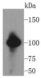 Western blot analysis of VAV2 on SW480 cells lysates using anti-VAV2 antibody at 1/1,000 dilution.
 -
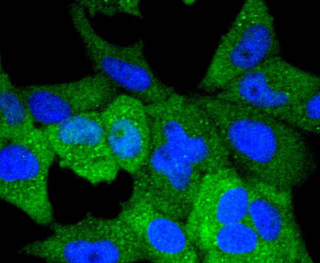 ICC staining VAV2 in Hela cells (green). The nuclear counter stain is DAPI (blue). Cells were fixed in paraformaldehyde, permeabilised with 0.25% Triton X100/PBS.
Bioworld Biotech only provide peptides for our antibodies and do not provide additional peptide customization services.
Price/Size :
USD 368/1mg/vial

Tips:
For phospho antibody, we provide phospho peptide(0.5mg) and non-phospho peptide(0.5mg).Describe :
Blocking peptides are peptides that bind specifically to the target antibody and block antibody binding. These peptide usually contains the epitope recognized by the antibody. Antibodies bound to the blocking peptide no longer bind to the epitope on the target protein. This mechanism is useful when non-specific binding is an issue, for example, in Western blotting (WB) and Immunohistochemistry (IHC). By comparing the staining from the blocked antibody versus the antibody alone, one can see which staining is specific; Specific binding will be absent from the western blot or IHC performed with the neutralized antibody.Formula:
Synthetic peptide was lyophilized with 100% acetonitrile and is supplied as a powder. Reconstitute with 0.1 ml DI water for a final concentration of 10 mg/ml.The purity is >90%,tested by HPLC and MS.
Storage:
The freeze-dried powder is more stable. For short time at 2-8°C. For long term storage store at -20°C.
Note :
This product is for research use only (RUO only). Not for use in diagnostic or therapeutic procedures.