
 villin1 polyclonal antibody
  Datasheet
 COA
COA MSDS
MSDS SHIP
SHIP
Product Name :
villin1 polyclonal antibody Background :
Caldesmon, Filamin 1, Nebulin and Villin are differentially expressed and regulated Actin binding proteins. Both muscular (CDh) and non-muscular (CDl) forms of Caldesmon have been identified and each has been shown to bind to Actin as well as to calmodulin and myosin. CDh is expressed predominantly on thin filaments in smooth muscle, whereas CDl is widely expressed in non-muscle tissues and cells. Filamin 1, which is ubiquitously expressed and exists as a homodimer, functions to crosslink Actin to filaments. Nebulin is a large filamentous protein specific to muscle tissue that may function as a ruler for filament length. Several isoforms of Nebulin are produced by alternative exon usage. Villin is Ca2+-regulated and is the major structural component of the brush border of absorptive cells. Product :
Rabbit IgG, 1mg/ml in PBS with 0.02% sodium azide, 50% glycerol, pH7.2 Storage&Stability :
Store at +4°C after thawing. Aliquot store at -20°C or -80°C. Avoid repeated freeze / thaw cycles. Specificity :
villin1 polyclonal antibody detects endogenous levels of villin1 protein. Immunogen :
Recombinant protein Conjugate :
Unconjugated Modification :
Unmodification
-
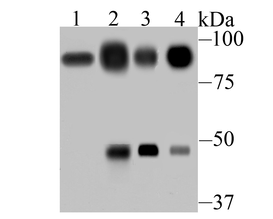 Western blot analysis of Villin1 on different tissue lysates using anti-Villin1 antibody at 1/500 dilution. Positive control: Lane 1: Mouse colon Lane 2: Human small intestine Lane 3: Human colon Lane 4: Rat kidney
 -
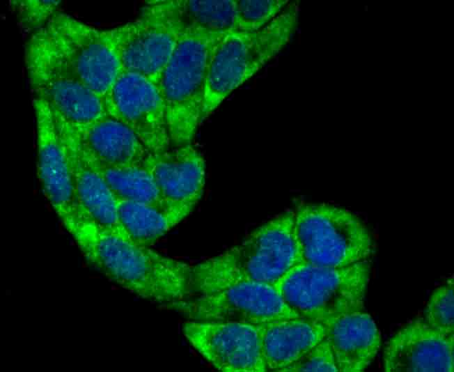 ICC staining Villin1 in Hela cells (green). The nuclear counter stain is DAPI (blue). Cells were fixed in paraformaldehyde, permeabilised with 0.25% Triton X100/PBS.
Bioworld Biotech only provide peptides for our antibodies and do not provide additional peptide customization services.
Price/Size :
USD 368/1mg/vial

Tips:
For phospho antibody, we provide phospho peptide(0.5mg) and non-phospho peptide(0.5mg).Describe :
Blocking peptides are peptides that bind specifically to the target antibody and block antibody binding. These peptide usually contains the epitope recognized by the antibody. Antibodies bound to the blocking peptide no longer bind to the epitope on the target protein. This mechanism is useful when non-specific binding is an issue, for example, in Western blotting (WB) and Immunohistochemistry (IHC). By comparing the staining from the blocked antibody versus the antibody alone, one can see which staining is specific; Specific binding will be absent from the western blot or IHC performed with the neutralized antibody.Formula:
Synthetic peptide was lyophilized with 100% acetonitrile and is supplied as a powder. Reconstitute with 0.1 ml DI water for a final concentration of 10 mg/ml.The purity is >90%,tested by HPLC and MS.
Storage:
The freeze-dried powder is more stable. For short time at 2-8°C. For long term storage store at -20°C.
Note :
This product is for research use only (RUO only). Not for use in diagnostic or therapeutic procedures.