
 EGFR (Phospho-S695) polyclonal antibody
  Datasheet
 COA
COA MSDS
MSDS SHIP
SHIP
Product Name :
EGFR (Phospho-S695) polyclonal antibody Background :
The EGF receptor family comprises several related receptor tyrosine kinases that are frequently overexpressed in a variety of carcinomas. Members of this receptor family include EGFR (HER1), Neu (ErbB-2, HER2), ErbB-3 (HER3) and ErbB-4 (HER4), which form either homodimers or heterodimers upon ligand binding. Exons in the EGFR gene product are frequently either deleted or duplicated to produce deletion mutants (DM) or tandem duplication mutants (TDM), respectively, which are detected at various molecular weights. EGFR binds several ligands, including epidermal growth factor (EGF), transforming growth factor α (TGFα), Amphiregulin and heparin binding-EGF (HB-EGF). Ligand binding promotes the internalization of EGFR via Clathrin-coated pits and its subsequent degradation in response to its intrinsic tyrosine kinase. EGFR is involved in organ morphogenesis and maintenance and repair of tissues, but upregulation of EGFR is associated with tumor progression. The oncogenic effects of EGFR include initiation of DNA synthesis, enhanced cell growth, invasion and metastasis. Abrogation of EGFR results in cell cycle arrest, apoptosis or dedifferentiation of cancer cells, suggesting that EGFR may be an effective therapeutic target. Product :
Rabbit IgG, 1mg/ml in PBS with 0.02% sodium azide, 50% glycerol, pH7.2 Storage&Stability :
Store at +4°C after thawing. Aliquot store at -20°C or -80°C. Avoid repeated freeze / thaw cycles. Specificity :
EGFR (Phospho-S695) polyclonal antibody detects endogenous levels of EGFR protein only when phosphorylated at S695. Immunogen :
Synthetic phospho-peptide corresponding to residues surrounding Ser695 of human EGFR. Conjugate :
Unconjugated Modification :
Phosphorylation
-
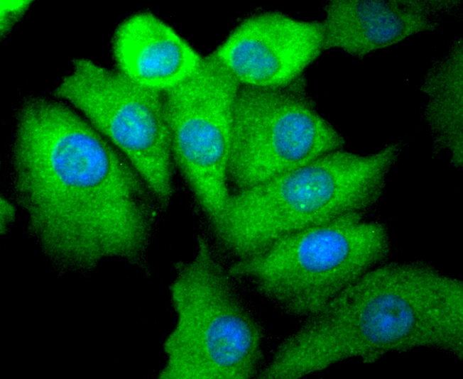 ICC staining Phospho-EGFR(S695) in A549 cells (green). The nuclear counter stain is DAPI (blue). Cells were fixed in paraformaldehyde, permeabilised with 0.25% Triton X100/PBS.
 -
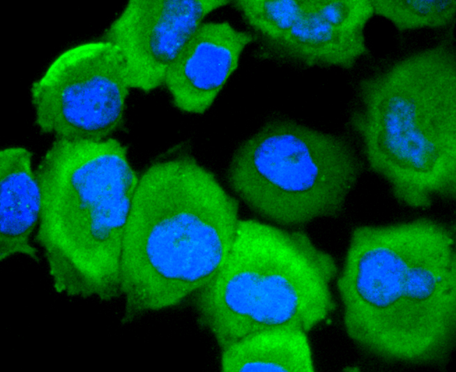 ICC staining Phospho-EGFR(S695) in HUVEC cells (green). The nuclear counter stain is DAPI (blue). Cells were fixed in paraformaldehyde, permeabilised with 0.25% Triton X100/PBS.
Bioworld Biotech only provide peptides for our antibodies and do not provide additional peptide customization services.
Price/Size :
USD 368/1mg/vial

Tips:
For phospho antibody, we provide phospho peptide(0.5mg) and non-phospho peptide(0.5mg).Describe :
Blocking peptides are peptides that bind specifically to the target antibody and block antibody binding. These peptide usually contains the epitope recognized by the antibody. Antibodies bound to the blocking peptide no longer bind to the epitope on the target protein. This mechanism is useful when non-specific binding is an issue, for example, in Western blotting (WB) and Immunohistochemistry (IHC). By comparing the staining from the blocked antibody versus the antibody alone, one can see which staining is specific; Specific binding will be absent from the western blot or IHC performed with the neutralized antibody.Formula:
Synthetic peptide was lyophilized with 100% acetonitrile and is supplied as a powder. Reconstitute with 0.1 ml DI water for a final concentration of 10 mg/ml.The purity is >90%,tested by HPLC and MS.
Storage:
The freeze-dried powder is more stable. For short time at 2-8°C. For long term storage store at -20°C.
Note :
This product is for research use only (RUO only). Not for use in diagnostic or therapeutic procedures.