
 c-Myc (Phospho-T58) polyclonal antibody
  Datasheet
 COA
COA MSDS
MSDS SHIP
SHIP
Product Name :
c-Myc (Phospho-T58) polyclonal antibody Background :
c-Myc-, N-Myc- and L-Myc-encoded proteins function in cell proliferation, differentiation and neoplastic disease. Myc proteins are nuclear proteins with relatively short half lives. Amplification of the c-Myc gene has been found in several types of human tumors including lung, breast and colon carcinomas, while the N-Myc gene has been found amplified in neuroblastomas. The L-Myc gene has been reported to be amplified and expressed at high level in human small cell lung carcinomas. The presence of three sequence motifs in the c-Myc COOH terminus, including the leucine zipper, the helix-loop-helix and a basic region provided initial evidence for a sequence-specific binding function. A basic region helix-loop-helix leucine zipper motif (bHLH-Zip) protein, designated Max, specifically associates with c-Myc, N-Myc and L-Myc proteins. The Myc-Max complex binds to DNA in a sequence-specific manner under conditions where neither Max nor Myc exhibit appreciable binding. Max can also form heterodimers with at least two additional bHLH-Zip proteins, Mad and Mxi1, and Mad-Max dimers have been shown to repress transcription through interaction with mSin3. Product :
Rabbit IgG, 1mg/ml in PBS with 0.02% sodium azide, 50% glycerol, pH7.2 Storage&Stability :
Store at +4°C after thawing. Aliquot store at -20°C or -80°C. Avoid repeated freeze / thaw cycles. Specificity :
c-Myc (Phospho-T58) polyclonal antibody detects endogenous levels of c-Myc protein only when phosphorylated at T58. Immunogen :
Synthetic phospho-peptide corresponding to residues surrounding Thr58 of human c-Myc Conjugate :
Unconjugated Modification :
Phosphorylation
-
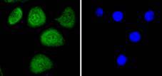
 ICC staining phospho-c-Myc(T58) in Hela cells (green). The nuclear counter stain is DAPI (blue). Cells were fixed in paraformaldehyde, permeabilised with 0.25% Triton X100/PBS.
 -
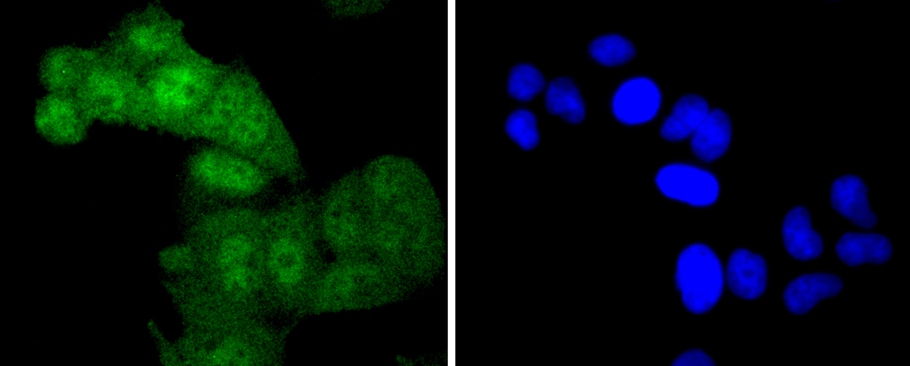
 ICC staining phospho-c-Myc(T58) in SKOV-3 cells (green). The nuclear counter stain is DAPI (blue). Cells were fixed in paraformaldehyde, permeabilised with 0.25% Triton X100/PBS.
Bioworld Biotech only provide peptides for our antibodies and do not provide additional peptide customization services.
Price/Size :
USD 368/1mg/vial

Tips:
For phospho antibody, we provide phospho peptide(0.5mg) and non-phospho peptide(0.5mg).Describe :
Blocking peptides are peptides that bind specifically to the target antibody and block antibody binding. These peptide usually contains the epitope recognized by the antibody. Antibodies bound to the blocking peptide no longer bind to the epitope on the target protein. This mechanism is useful when non-specific binding is an issue, for example, in Western blotting (WB) and Immunohistochemistry (IHC). By comparing the staining from the blocked antibody versus the antibody alone, one can see which staining is specific; Specific binding will be absent from the western blot or IHC performed with the neutralized antibody.Formula:
Synthetic peptide was lyophilized with 100% acetonitrile and is supplied as a powder. Reconstitute with 0.1 ml DI water for a final concentration of 10 mg/ml.The purity is >90%,tested by HPLC and MS.
Storage:
The freeze-dried powder is more stable. For short time at 2-8°C. For long term storage store at -20°C.
Note :
This product is for research use only (RUO only). Not for use in diagnostic or therapeutic procedures.