
 Histone H1.3/H1.4 (phospho-T17) Rabbit monoclonal antibody
  Datasheet
 COA
COA MSDS
MSDS SHIP
SHIP
Product Name :
Histone H1.3/H1.4 (phospho-T17) Rabbit monoclonal antibody Background :
Eukaryotic histones are basic and water soluble nuclear proteins that form hetero-octameric nucleosome particles by wrapping 146 base pairs of DNA in a left-handed super-helical turn sequentially to form chromosomal fiber. Two molecules of each of the four core histones (H2A, H2B, H3, and H4) form the octamer; formed of two H2A-H2B dimers and two H3-H4 dimers, forming two nearly symmetrical halves by tertiary structure. Over 80% of nucleosomes contain the linker Histone H1, derived from an intronless gene, that interacts with linker DNA between nucleosomes and mediates compaction into higher order chromatin. Histones are subject to posttranslational modification by enzymes primarily on their N-terminal tails, but also in their globular domains. Such modifications include methylation, citrullination, acetylation, phosphorylation, sumoylation, ubiquitination and ADP-ribosylation. Product :
Rabbit IgG, 1mg/ml in PBS with 0.02% sodium azide, 50% glycerol, pH7.2 Storage&Stability :
Store at 4°C short term. Aliquot and store at -20°C long term. Avoid freeze-thaw cycles. Specificity :
This antibody detects endogenous levels of Histone H1.3 or H1.4 protein only when pho17. Immunogen :
Recombinant antibody. Conjugate :
Unconjugated Modification :
Phosphorylation
-
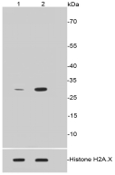 Western blot (WB) analysis of Histone H1.3/H1.4 (phospho-T17) Rabbit mAb at 1:500 dilution Lane 1: Untreated CRC whole cell lysates Lane2: CRC cells treated with 1.5µg/ml Colcemid for 12 hours whole cell lysates
 -
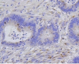 Immunohistochemical analysis of paraffin-embedded human colon cancer tissue using anti-Histone H1.3/H1.4 (phospho-T17) antibody. Counter stained with hematoxylin.
 -
 Immunohistochemical analysis of paraffin-embedded human colon cancer tissue using anti-Histone H1.3/H1.4 (phospho-T17) antibody. Counter stained with hematoxylin.
Bioworld Biotech only provide peptides for our antibodies and do not provide additional peptide customization services.
Price/Size :
USD 368/1mg/vial

Tips:
For phospho antibody, we provide phospho peptide(0.5mg) and non-phospho peptide(0.5mg).Describe :
Blocking peptides are peptides that bind specifically to the target antibody and block antibody binding. These peptide usually contains the epitope recognized by the antibody. Antibodies bound to the blocking peptide no longer bind to the epitope on the target protein. This mechanism is useful when non-specific binding is an issue, for example, in Western blotting (WB) and Immunohistochemistry (IHC). By comparing the staining from the blocked antibody versus the antibody alone, one can see which staining is specific; Specific binding will be absent from the western blot or IHC performed with the neutralized antibody.Formula:
Synthetic peptide was lyophilized with 100% acetonitrile and is supplied as a powder. Reconstitute with 0.1 ml DI water for a final concentration of 10 mg/ml.The purity is >90%,tested by HPLC and MS.
Storage:
The freeze-dried powder is more stable. For short time at 2-8°C. For long term storage store at -20°C.
Note :
This product is for research use only (RUO only). Not for use in diagnostic or therapeutic procedures.