
 FGFR2/CD332 Mouse monoclonal antibody
  Datasheet
 COA
COA MSDS
MSDS SHIP
SHIP
Product Name :
FGFR2/CD332 Mouse monoclonal antibody Background :
Fibroblast growth factor receptor 2 (FGFR2) also known as CD332 has two naturally occurring isoforms FGFR2IIIb and FGFR2IIIc, created by splicing of the third immunoglobulin-like domain. FGFR2IIIb is predominantly found in ectoderm derived tissues and endothelial organ lining, i.e. skin and internal organs. FGFR2 has important roles in embryonic development and tissue repair, especially bone and blood vessels. Like the other members of the Fibroblast growth factor receptor family, these receptors signal by binding to their ligand and dimerisation (pairing of receptors), which causes the tyrosine kinase domains to initiate a cascade of intracellular signals. As mentioned, FGFR2 mutations are associated with craniosynostosis syndromes, which are skull malformations caused by premature fusion of cranial sutures and other disease features according to the mutation itself. Product :
Mouse IgG1, 1mg/ml in PBS with 0.02% sodium azide, 50% glycerol, pH7.2 Storage&Stability :
Store at 4°C short term. Aliquot and store at -20°C long term. Avoid freeze-thaw cycles. Specificity :
This antibody detects endogenous levels of FGFR2 and does not cross-react with related proteins. Immunogen :
This antibody is produced by immunizing mice with a recombinant protein corresponding to a region of FGFR2. Conjugate :
Unconjugated Modification :
Unmodification
-
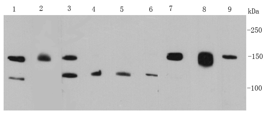 Western blot (WB) analysis of FGFR2 Mouse mAb at 1:500 dilution Lane1:MCF7 whole cell lysate Lane2:K562 whole cell lysate Lane3:Hela whole cell lysate Lane4:HepG2 whole cell lysate Lane5:A431 whole cell lysate Lane6:A549 whole cell lysate Lane7:NIH/3T3 whole cell lysate Lane8:Jurkat whole cell lysate
 -
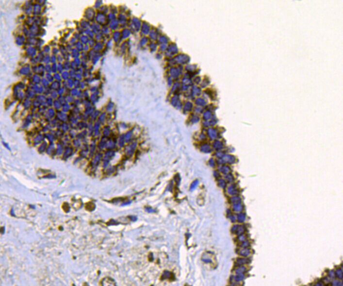 Immunohistochemical analysis of paraffin-embedded human breast carcinoma tissue using anti-FGFR2 antibody. Counter stained with hematoxylin.
 -
 Immunohistochemical analysis of paraffin-embedded human breast carcinoma tissue using anti-FGFR2 antibody. Counter stained with hematoxylin.
Bioworld Biotech only provide peptides for our antibodies and do not provide additional peptide customization services.
Price/Size :
USD 368/1mg/vial

Tips:
For phospho antibody, we provide phospho peptide(0.5mg) and non-phospho peptide(0.5mg).Describe :
Blocking peptides are peptides that bind specifically to the target antibody and block antibody binding. These peptide usually contains the epitope recognized by the antibody. Antibodies bound to the blocking peptide no longer bind to the epitope on the target protein. This mechanism is useful when non-specific binding is an issue, for example, in Western blotting (WB) and Immunohistochemistry (IHC). By comparing the staining from the blocked antibody versus the antibody alone, one can see which staining is specific; Specific binding will be absent from the western blot or IHC performed with the neutralized antibody.Formula:
Synthetic peptide was lyophilized with 100% acetonitrile and is supplied as a powder. Reconstitute with 0.1 ml DI water for a final concentration of 10 mg/ml.The purity is >90%,tested by HPLC and MS.
Storage:
The freeze-dried powder is more stable. For short time at 2-8°C. For long term storage store at -20°C.
Note :
This product is for research use only (RUO only). Not for use in diagnostic or therapeutic procedures.