
 APE1 monoclonal antibody
  Datasheet
 COA
COA MSDS
MSDS SHIP
SHIP
Product Name :
APE1 monoclonal antibody Background :
The role of transcription factors in the regulation of gene expression is well established. Although the activity of these factors can be regulated by phosphorylation, evidence has indicated regulation of DNA binding mediated by changes in reduction-oxidation (redox) status. Mutational analysis has identified a single conserved cysteine residue mapping within the DNA binding domains of Fos and Jun. Chemical oxidation or modification of this cysteine residue inhibits the DNA binding activity of Fos and Jun. A similar mode of regulation has been recently proposed for other nuclear transcription factors. Oxidation is reversible by these compounds or by a cellular redox/DNA repair protein identified originally as Ref-1 (redox factor 1). Ref-1 is identical to a previously characterized DNA repair enzyme designated HAP1, APE or APEX. Product :
Mouse IgG2a, 1mg/ml in PBS with 0.02% sodium azide, 50% glycerol, pH7.2 Storage&Stability :
Store at +4℃ after thawing. Aliquot store at -20℃. Avoid repeated freeze / thaw cycles. Specificity :
APE1 monoclonal antibody detects endogenous levels of APE1 protein. Immunogen :
Recombinant protein within Human APE1 aa 20-350. Conjugate :
Unconjugated Modification :
Unmodification
-
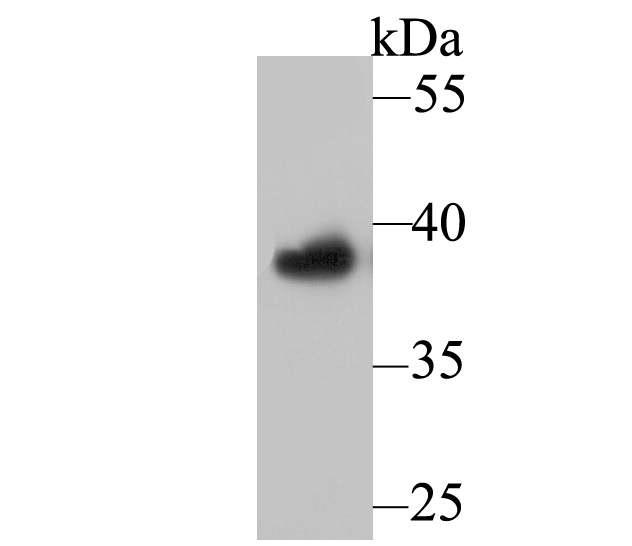 Western blot analysis of APE1 on HL-60 lysates. Proteins were transferred to a PVDF membrane and blocked with 5% BSA in PBS for 1 hour at room temperature. The primary antibody was used at a 1:1,000 dilution in 5% BSA at room temperature for 2 hours. Goat Anti-Mouse IgG - HRP Secondary Antibody (HA1006) at 1:5,000 dilution was used for 1 hour at room temperature.
 -
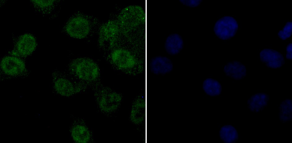 ICC staining APE1 in A549 cells (green). Formalin fixed cells were permeabilized with 0.1% Triton X-100 in TBS for 10 minutes at room temperature and blocked with 1% Blocker BSA for 15 minutes at room temperature. Cells were probed with APE1 monoclonal antibody at a dilution of 1:50 for 1 hour at room temperature, washed with PBS. Alexa Fluorc™ 488 Goat anti-Mouse IgG was used as the secondary antibody at 1/100 dilution. The nuclear counter stain is DAPI (blue).
Bioworld Biotech only provide peptides for our antibodies and do not provide additional peptide customization services.
Price/Size :
USD 368/1mg/vial

Tips:
For phospho antibody, we provide phospho peptide(0.5mg) and non-phospho peptide(0.5mg).Describe :
Blocking peptides are peptides that bind specifically to the target antibody and block antibody binding. These peptide usually contains the epitope recognized by the antibody. Antibodies bound to the blocking peptide no longer bind to the epitope on the target protein. This mechanism is useful when non-specific binding is an issue, for example, in Western blotting (WB) and Immunohistochemistry (IHC). By comparing the staining from the blocked antibody versus the antibody alone, one can see which staining is specific; Specific binding will be absent from the western blot or IHC performed with the neutralized antibody.Formula:
Synthetic peptide was lyophilized with 100% acetonitrile and is supplied as a powder. Reconstitute with 0.1 ml DI water for a final concentration of 10 mg/ml.The purity is >90%,tested by HPLC and MS.
Storage:
The freeze-dried powder is more stable. For short time at 2-8°C. For long term storage store at -20°C.
Note :
This product is for research use only (RUO only). Not for use in diagnostic or therapeutic procedures.