
 Ribonuclease 3 monoclonal antibody
  Datasheet
 COA
COA MSDS
MSDS SHIP
SHIP
Product Name :
Ribonuclease 3 monoclonal antibody Background :
The protein encoded by this gene belongs to the pancreatic ribonuclease family, a subset of the ribonuclease A superfamily. Cytotoxin and helminthotoxin with low-efficiency ribonuclease activity. The protein exhibits antimicrobial activity against pathogenic bacteria. Exhibits antibacterial activity, including cytoplasmic membrane depolarization of preferentially Gram-negative, but also Gram-positive strains. Promotes E.coli outer membrane detachment, alteration of the overall cell shape and partial loss of cell content. ECP is a potent cytotoxic protein capable of killing cells of guinea pig tracheal epithelium, mammalian leukemia, epidermis carcinoma, and breast carcinoma, as well as non-mammalian cells such as parasites, bacteria, and viruses. Mature ECP is cytotoxic to human bronchial epithelial (BEAS-2B) cells by specific binding to cell surface heparan sulfate proteoglycans (HSPGs) followed by endocytosis. ECP triggers apoptosis by caspase-8 activation through mitochondria-independent pathway. Increases in chromatin condensation, sub-G1 population, PARP cleavage, and DNA fragmentation indicate that ECP induces apoptosis in human bronchial epithelial (BEAS-2B) cells. Product :
Mouse IgG1, 1mg/ml in PBS with 0.02% sodium azide, 50% glycerol, pH7.2 Storage&Stability :
Store at +4℃ after thawing. Aliquot store at -20℃. Avoid repeated freeze / thaw cycles. Specificity :
Ribonuclease 3 monoclonal antibody detects endogenous levels of Ribonuclease 3 protein. Immunogen :
Recombinant protein within human Ribonuclease 3 aa 28-120. Conjugate :
Unconjugated Modification :
Unmodification
-
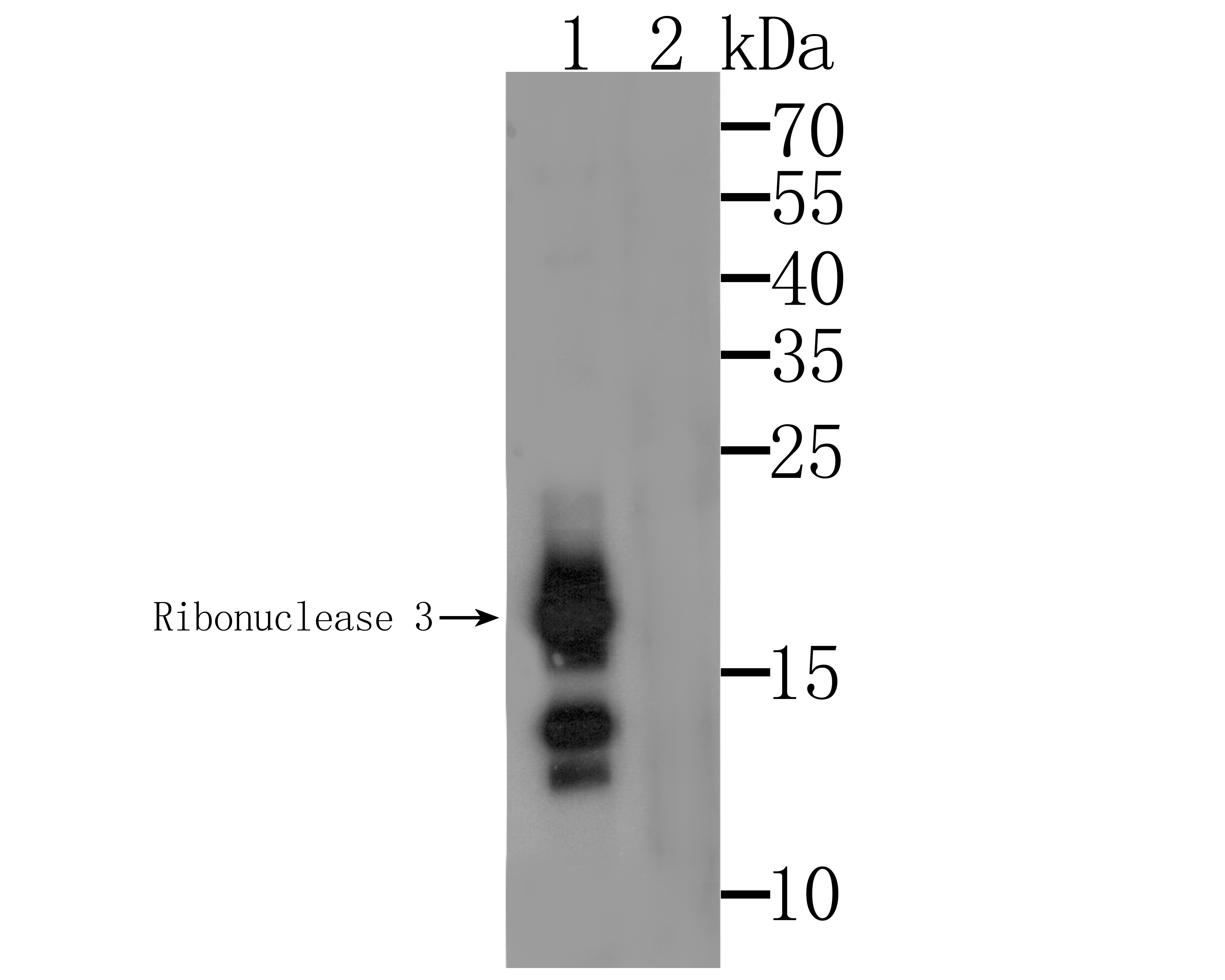 Western blot analysis of Ribonuclease 3 on U937 cell lysate. Proteins were transferred to a PVDF membrane and blocked with 5% BSA in PBS for 1 hour at room temperature. The primary antibody was used at a 1:500 dilution in 5% BSA at room temperature for 2 hours. Goat Anti-Mouse IgG - HRP Secondary Antibody (HA1006) at 1:5,000 dilution was used for 1 hour at room temperature.Lane 1: Anti-Ribonuclease 3 Antibody, (1:500).Lane 2: Anti-Ribonuclease 3 Antibody, (1:500), preincubated with the immunizat
 -
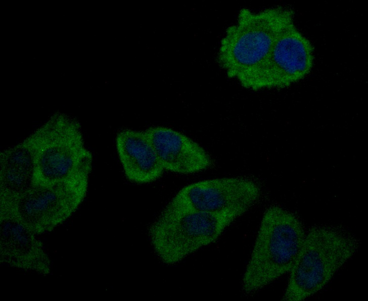 ICC staining Ribonuclease 3 in Hela cells (green). Formalin fixed cells were permeabilized with 0.1% Triton X-100 in TBS for 10 minutes at room temperature and blocked with 1% Blocker BSA for 15 minutes at room temperature. Cells were probed with Ribonuclease 3 monoclonal antibody at a dilution of 1:50 for 1 hour at room temperature, washed with PBS. Alexa Fluorc™ 488 Goat anti-Mouse IgG was used as the secondary antibody at 1/100 dilution. The nuclear counter stain is DAPI (blue).
Bioworld Biotech only provide peptides for our antibodies and do not provide additional peptide customization services.
Price/Size :
USD 368/1mg/vial

Tips:
For phospho antibody, we provide phospho peptide(0.5mg) and non-phospho peptide(0.5mg).Describe :
Blocking peptides are peptides that bind specifically to the target antibody and block antibody binding. These peptide usually contains the epitope recognized by the antibody. Antibodies bound to the blocking peptide no longer bind to the epitope on the target protein. This mechanism is useful when non-specific binding is an issue, for example, in Western blotting (WB) and Immunohistochemistry (IHC). By comparing the staining from the blocked antibody versus the antibody alone, one can see which staining is specific; Specific binding will be absent from the western blot or IHC performed with the neutralized antibody.Formula:
Synthetic peptide was lyophilized with 100% acetonitrile and is supplied as a powder. Reconstitute with 0.1 ml DI water for a final concentration of 10 mg/ml.The purity is >90%,tested by HPLC and MS.
Storage:
The freeze-dried powder is more stable. For short time at 2-8°C. For long term storage store at -20°C.
Note :
This product is for research use only (RUO only). Not for use in diagnostic or therapeutic procedures.