
 N-WASP/WASL polyclonal antibody
  Datasheet
 COA
COA MSDS
MSDS SHIP
SHIP
Product Name :
N-WASP/WASL polyclonal antibody Background :
This gene encodes a member of the Wiskott-Aldrich syndrome (WAS) protein family. Wiskott-Aldrich syndrome proteins share similar domain structure, and associate with a variety of signaling molecules to alter the actin cytoskeleton. The encoded protein is highly expressed in neural tissues, and interacts with several proteins involved in cytoskeletal organization, including cell division control protein 42 (CDC42) and the actin-related protein-2/3 (ARP2/3) complex. The encoded protein may be involved in the formation of long actin microspikes, and in neurite extension. Product :
1mg/ml in PBS with 0.02% sodium azide, 50% glycerol, pH7.2 Storage&Stability :
Store at 4°C short term. Aliquot and store at -20°C long term. Avoid freeze-thaw cycles. Specificity :
Polyclonal Antibodies Immunogen :
Recombinant fusion protein of human N-WASP/WASL(NP_003932.3). Conjugate :
Unconjugated Modification :
Unmodification
-
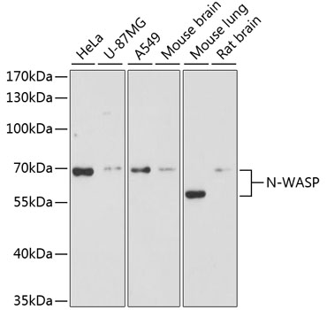 Western blot analysis of extracts of various cell lines, using N-WASP/WASL antibody at 1:3000 dilution.
Secondary antibody: HRP Goat Anti-Rabbit IgG at 1:10000 dilution.
Lysates/proteins: 25ug per lane.
Blocking buffer: 3% nonfat dry milk in TBST.
Detection: ECL Basic Kit .
Exposure time: 90s. -
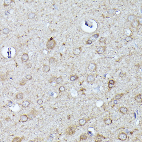 Immunohistochemistry of paraffin-embedded rat brain using N-WASP/WASL antibody at dilution of 1:100 .Perform microwave antigen retrieval with 10 mM PBS buffer pH 7.2 before commencing with IHC staining protocol.
 -
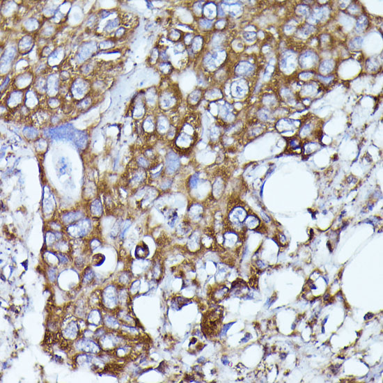 Immunohistochemistry of paraffin-embedded rat brain using N-WASP/WASL antibody at dilution of 1:100 .Perform microwave antigen retrieval with 10 mM PBS buffer pH 7.2 before commencing with IHC staining protocol.
 -
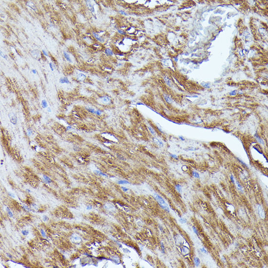 Immunohistochemistry of paraffin-embedded rat brain using N-WASP/WASL antibody at dilution of 1:100 .Perform microwave antigen retrieval with 10 mM PBS buffer pH 7.2 before commencing with IHC staining protocol.
Bioworld Biotech only provide peptides for our antibodies and do not provide additional peptide customization services.
Price/Size :
USD 368/1mg/vial

Tips:
For phospho antibody, we provide phospho peptide(0.5mg) and non-phospho peptide(0.5mg).Describe :
Blocking peptides are peptides that bind specifically to the target antibody and block antibody binding. These peptide usually contains the epitope recognized by the antibody. Antibodies bound to the blocking peptide no longer bind to the epitope on the target protein. This mechanism is useful when non-specific binding is an issue, for example, in Western blotting (WB) and Immunohistochemistry (IHC). By comparing the staining from the blocked antibody versus the antibody alone, one can see which staining is specific; Specific binding will be absent from the western blot or IHC performed with the neutralized antibody.Formula:
Synthetic peptide was lyophilized with 100% acetonitrile and is supplied as a powder. Reconstitute with 0.1 ml DI water for a final concentration of 10 mg/ml.The purity is >90%,tested by HPLC and MS.
Storage:
The freeze-dried powder is more stable. For short time at 2-8°C. For long term storage store at -20°C.
Note :
This product is for research use only (RUO only). Not for use in diagnostic or therapeutic procedures.