
 PDP1 polyclonal antibody
  Datasheet
 COA
COA MSDS
MSDS SHIP
SHIP
Product Name :
PDP1 polyclonal antibody Background :
Pyruvate dehydrogenase (E1) is one of the three components (E1, E2, and E3) of the large pyruvate dehydrogenase complex. Pyruvate dehydrogenase kinases catalyze phosphorylation of serine residues of E1 to inactivate the E1 component and inhibit the complex. Pyruvate dehydrogenase phosphatases catalyze the dephosphorylation and activation of the E1 component to reverse the effects of pyruvate dehydrogenase kinases. Pyruvate dehydrogenase phosphatase is a heterodimer consisting of catalytic and regulatory subunits. Two catalytic subunits have been reported; one is predominantly expressed in skeletal muscle and another one is is much more abundant in the liver. The catalytic subunit, encoded by this gene, is the former, and belongs to the protein phosphatase 2C (PP2C) superfamily. Along with the pyruvate dehydrogenase complex and pyruvate dehydrogenase kinases, this enzyme is located in the mitochondrial matrix. Mutation in this gene causes pyruvate dehydrogenase phosphatase deficiency. Multiple alternatively spliced transcript variants encoding different isoforms have been identified. Product :
1mg/ml in PBS with 0.02% sodium azide, 50% glycerol, pH7.2 Storage&Stability :
Store at 4°C short term. Aliquot and store at -20°C long term. Avoid freeze-thaw cycles. Specificity :
Polyclonal Antibodies Immunogen :
Recombinant fusion protein of human PDP1(NP_060914.2). Conjugate :
Unconjugated Modification :
Unmodification
-
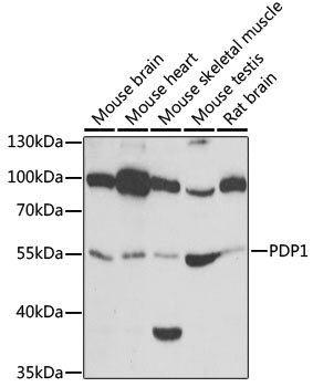 Western blot analysis of extracts of various cell lines, using PDP1 antibody at 1:1000 dilution.
Secondary antibody: HRP Goat Anti-Rabbit IgG at 1:10000 dilution.
Lysates/proteins: 25ug per lane.
Blocking buffer: 3% nonfat dry milk in TBST.
Detection: ECL Basic Kit .
Exposure time: 30s. -
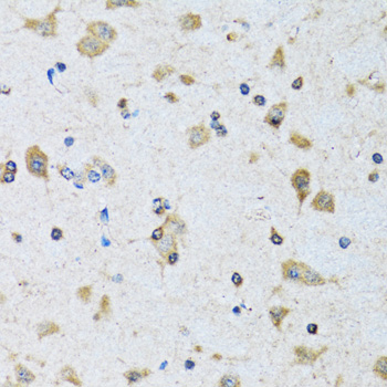 Immunohistochemistry of paraffin-embedded rat brain using PDP1 antibody at dilution of 1:100 .Perform microwave antigen retrieval with 10 mM PBS buffer pH 7.2 before commencing with IHC staining protocol.
 -
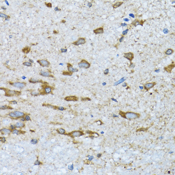 Immunohistochemistry of paraffin-embedded rat brain using PDP1 antibody at dilution of 1:100 .Perform microwave antigen retrieval with 10 mM PBS buffer pH 7.2 before commencing with IHC staining protocol.
 -
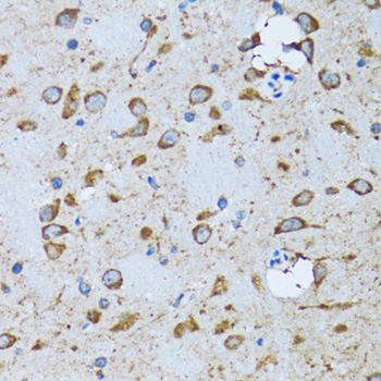 Immunohistochemistry of paraffin-embedded rat brain using PDP1 antibody at dilution of 1:100 .Perform microwave antigen retrieval with 10 mM PBS buffer pH 7.2 before commencing with IHC staining protocol.
Bioworld Biotech only provide peptides for our antibodies and do not provide additional peptide customization services.
Price/Size :
USD 368/1mg/vial

Tips:
For phospho antibody, we provide phospho peptide(0.5mg) and non-phospho peptide(0.5mg).Describe :
Blocking peptides are peptides that bind specifically to the target antibody and block antibody binding. These peptide usually contains the epitope recognized by the antibody. Antibodies bound to the blocking peptide no longer bind to the epitope on the target protein. This mechanism is useful when non-specific binding is an issue, for example, in Western blotting (WB) and Immunohistochemistry (IHC). By comparing the staining from the blocked antibody versus the antibody alone, one can see which staining is specific; Specific binding will be absent from the western blot or IHC performed with the neutralized antibody.Formula:
Synthetic peptide was lyophilized with 100% acetonitrile and is supplied as a powder. Reconstitute with 0.1 ml DI water for a final concentration of 10 mg/ml.The purity is >90%,tested by HPLC and MS.
Storage:
The freeze-dried powder is more stable. For short time at 2-8°C. For long term storage store at -20°C.
Note :
This product is for research use only (RUO only). Not for use in diagnostic or therapeutic procedures.