
 RIPK1/RIP polyclonal antibody
  Datasheet
 COA
COA MSDS
MSDS SHIP
SHIP
Product Name :
RIPK1/RIP polyclonal antibody Background :
3'-5' DNA helicase and substrate-recognition component of the SCF(FBH1 E3 ubiquitin ligase complex that plays a key role in response to stalled/damaged replication forks. Involved in genome maintenance by acting as an anti-recombinogenic helicase and preventing extensive strand exchange during homologous recombination: promotes RAD51 filament dissolution from stalled forks, thereby inhibiting homologous recombination and preventing excessive recombination. Also promotes cell death and DNA double-strand breakage in response to replication stress: together with MUS81, promotes the endonucleolytic DNA cleavage following prolonged replication stress via its helicase activity, possibly to eliminate cells with excessive replication stress. Plays a major role in remodeling of stalled DNA forks by catalyzing fork regression, in which the fork reverses and the two nascent DNA strands anneal. In addition to the helicase activity, also acts as the substrate-recognition component of the SCF(FBH1 E3 ubiquitin ligase complex, a complex that mediates ubiquitination of RAD51, leading to regulate RAD51 subcellular location. Product :
1mg/ml in PBS with 0.02% sodium azide, 50% glycerol, pH7.2 Storage&Stability :
Store at 4°C short term. Aliquot and store at -20°C long term. Avoid freeze-thaw cycles. Specificity :
RIPK1/RIP polyclonal antibody detects endogenous levels of RIPK1/RIP protein. Immunogen :
A synthetic peptide corresponding to a sequence within amino acids 100-200 of human RIPK1/RIP (NP_003795.2). Conjugate :
Unconjugated Modification :
Unmodified
-
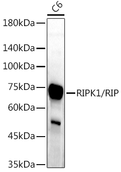 Western blot analysis of extracts of C6 cells, using RIPK1/RIP antibody at 1:1000 dilution.
Secondary antibody: HRP Goat Anti-Rabbit IgG at 1:10000 dilution.
Lysates/proteins: 25ug per lane.
Blocking buffer: 3% nonfat dry milk in TBST.
Detection: ECL Enhanced Kit .
Exposure time: 180s. -
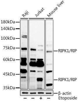 Western blot analysis of extracts of various cell lines, using RIPK1/RIP antibody at 1:1000 dilution.Jurkat cells were treated by Etoposide at 37℃ for 5 hours.
Secondary antibody: HRP Goat Anti-Rabbit IgG at 1:10000 dilution.
Lysates/proteins: 25ug per lane.
Blocking buffer: 3% nonfat dry milk in TBST.
Detection: ECL Basic Kit .
Exposure time: 90s. -
 Western blot analysis of extracts of various cell lines, using RIPK1/RIP antibody at 1:1000 dilution.Jurkat cells were treated by Etoposide at 37℃ for 5 hours.
Secondary antibody: HRP Goat Anti-Rabbit IgG at 1:10000 dilution.
Lysates/proteins: 25ug per lane.
Blocking buffer: 3% nonfat dry milk in TBST.
Detection: ECL Basic Kit .
Exposure time: 90s.
Bioworld Biotech only provide peptides for our antibodies and do not provide additional peptide customization services.
Price/Size :
USD 368/1mg/vial

Tips:
For phospho antibody, we provide phospho peptide(0.5mg) and non-phospho peptide(0.5mg).Describe :
Blocking peptides are peptides that bind specifically to the target antibody and block antibody binding. These peptide usually contains the epitope recognized by the antibody. Antibodies bound to the blocking peptide no longer bind to the epitope on the target protein. This mechanism is useful when non-specific binding is an issue, for example, in Western blotting (WB) and Immunohistochemistry (IHC). By comparing the staining from the blocked antibody versus the antibody alone, one can see which staining is specific; Specific binding will be absent from the western blot or IHC performed with the neutralized antibody.Formula:
Synthetic peptide was lyophilized with 100% acetonitrile and is supplied as a powder. Reconstitute with 0.1 ml DI water for a final concentration of 10 mg/ml.The purity is >90%,tested by HPLC and MS.
Storage:
The freeze-dried powder is more stable. For short time at 2-8°C. For long term storage store at -20°C.
Note :
This product is for research use only (RUO only). Not for use in diagnostic or therapeutic procedures.